One major limitation associated with traditional optical microscopy imaging of tissues is the inability to image beyond a few hundred micrometers in the visible spectrum or beyond the range of one to 1.5 millimeters (mm) in the infrared spectrum.
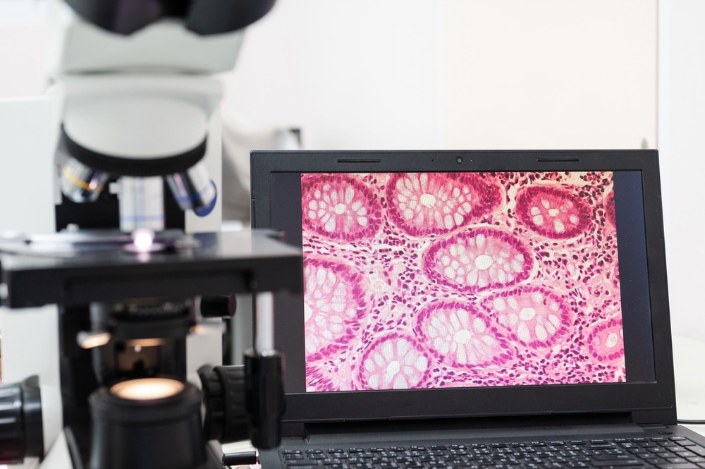
Image Credit: Komsan Loonprom/Shutterstock.com
As a result of these limitations, researchers have looked at alternative approaches to optical microscopy, such as optical-clearing methods, that could improve their tissue-penetration depth. These methods require the chemical processing of samples to allow them to become transparent; however, these methods are often toxic to tissues and can therefore only be used in post-mortem samples.
What is Mesoscopy?
To overcome the limitations associated with optical microscopy when deep tissue visualization is required, optoacoustic mesoscopy (OPAM) has been proposed as a bridge between optoacoustic macroscopy and optoacoustic microscopy. OPAM imaging, which was first considered in the 1970s, allows in vivo imaging with a tissue-penetration depth reaching up to several centimeters (cm).
Because OPAM imaging is insensitive to photon scatter, this technique can image deep inside tissues by resolving the absorption of light by detecting ultrasound waves rather than photons. Within the live tissue sample, ultrasound waves are generated in response to the thermoelastic expansion of tissue moieties that absorb transient light energy. Thus, the role of ultrasound diffraction in forming the final image is why this method is often referred to as acoustic-resolution (AR) optoacoustic imaging.
Optoacoustic macroscopy, which allows the user to visualize several centimeters into the tissue, also uses ultrasound arrays to achieve resolutions of several hundreds of micrometers (µm). At these frequencies, whole-body animal imaging is possible and has also been used in humans for the non-invasive imaging of breast cancer, intestinal inflammation, and brown-fat metabolism.
Compared to optoacoustic macroscopy, OPAM involves much higher ultrasound frequencies that subsequently allow the tissue-penetration depth to reach a few mm, all the while retaining resolutions that are typically within the 30-100 µm range.
Applications of Meoscopy
Several subtypes of OPAM have been developed for biomedical applications. These imaging techniques often allow for the non-invasive imaging and deep penetration of tissues without compromising the resolution of the final images. As a result of its tissue-penetration depth capabilities, OPAM methods can provide important information on the structural, functional, and molecular features of a wide range of medical diseases.
Image Credit: Africa Studio/Shutterstock.com
Dermatology
Within the field of dermatology, raster-scan OPAM (RSOM) has emerged as a particularly useful imaging tool for diagnosing and managing several dermatological conditions.
One of the key advantages of RSOM and other OPAM techniques in diagnosing dermatological conditions is that it overcomes some of the traditional challenges associated with the visual inspection of skin conditions. Although a formal skin condition diagnosis is often accompanied by histopathological analysis of the affected skin, this method is invasive and associated with an increased risk of bleeding, infection, and scar formation.
In particular, RSOM is increasingly widely used to assess inflammatory skin conditions like psoriasis and eczema, as it allows for more accurate quantification of inflammation-related alterations within the skin. Psoriasis, for example, is often associated with increased vasculature and thickness of the epidermis, can successfully be visualized with single wavelength RSOM, and provide observations that correlate with histological imaging of psoriasis patients.
Furthermore, when atopic dermatitis is present, the inflammation of this condition will cause dermal vasculature changes that often include increased dilation and vessel density. Taken together, these characteristics of atopic dermatitis can be visualized with RSOM, thereby eliminating the need for skin biopsies to diagnose this condition.
As this diagnostic technique continues to advance, researchers are finding that RSOM can also be used to quantify the presence of inflammatory markers within the skin. These types of refinements in RSOM and other OPAM techniques will not only ensure the accurate diagnosis of skin conditions but will also allow dermatologists to monitor the progress of patients following the prescription of various topical agents. If the levels of certain biomarkers are unchanged and/or worsen, clinicians can quickly alter their treatment of choice to serve each patient's specific needs better.
Gastroenterology
Several research efforts have also looked to RSOM to enhance the sensitivity of current imaging technologies used in gastroenterology. For example, one recent study integrated RSOM into a small animal scanner, wherein the researchers obtained label-free images of freshly excised murine colons that were both inflamed and contained tumors.
To this end, the researchers found that their system successfully visualized the increased vasculature within the colon wall and compared these images to that of a normal and healthy mouse colon. Furthermore, RSOM successfully detected structures that resembled labeled T-cells in a T-cell transfer colitis mouse model.
Although the application of RSOM and other OPAM methods are still in the early stages of development for gastroenterology diagnoses, researchers anticipate that this imaging modality will significantly improve the resolution of current optical imaging technologies. The integration of RSOM into endoscopic cameras, for example, could greater increase the depth of intestinal wall imaging to provide more accurate diagnoses of upper gastrointestinal diseases without needing to obtain biopsies of diseased areas.
Sources:
- Omar, M., Aguirre, J., & Ntziachristos, B. (2019). Optoacoustic mesoscopy for biomedicine. Nature Biomedical Engineering 3; 354-370. doi:10.1038/s41551-019-0377-4.
- Messas, T., Messas, A., & Kroumpouzos, G. (2021). Optoacoustic Imaging And Potential Applications of Raster-Scan Optoacoustic Mesoscopy in Dermatology. Clinics in Dermatology. doi:10.1016/j.clindermatol.2021.12.001.
- Knieling, F., Menezes, J. G., Claussen, J., et al. (2018). Raster-Scanning Optoacoustic Mesoscopy for Gastrointestinal Imaging at High Resolution. Gastroenterology in Motion 154(4); 807-809. doi:10.1053/j.gastro.2017.11.285.
Further Reading
Last Updated: Jun 29, 2022
 Laser Phase Plate in the Electron Microscope Promises Clear Images of Tiniest Proteins