The microscope allowed us to view living biological systems for the very first time. Systems that were inaccessible to the naked eye were suddenly available to scientists, allowing them to confirm or deny theories on the workings of life. Since its invention in the late 16th century, the microscope has been at the center of numerous scientific breakthroughs, particularly in biology, facilitating discoveries and confirming hypotheses that have helped vastly develop our understanding of humans, animals, and plants.
Now, as digitalization sweeps across all sectors, radically changing them forever, the field of microscopy is also heading into a new era. In the last few decades, digital microscopy has become more widely adopted and is being used over traditional microscopy for its many benefits. Here, we outline the evolution of the microscope, from the first microscope created in 1590 to the birth of the digital microscope in 1986. We consider how the field may continue evolving, highlighting some key manufacturers.
A Brief History Of The Microscope
The evolution of traditional microscopy
The microscope was invented by Hans and Zacharias Janssen, a Dutch father and son team who worked as spectacle makers in 1590. This invention led to the publication of the groundbreaking book "Micrographia" in 1667, authored by Robert Hooke. The book contains large-scale and incredibly detailed illustrations of the specimens that Hooke examined through the microscope. It is considered to be the first important work of microscopy. Hooke's detailed drawings and observations helped other scientists understand biology at this fine-grain level that had never been seen before.
Less than a decade later, Anton van Leeuwenhoek became the first person to ever observe bacteria. This marked the beginning of a new era, and microscopy became widely popular among scientists throughout the following century.
By the 19th century, the design of microscopes was becoming more sophisticated, with Joseph Jackson Lister's discovery of increasing the clarity of magnification by combining weak lenses together at various distances, particularly fueling this evolution.
By the 20th century, the field of microscopy was evolving faster than ever. In 1903, the ultramicroscope was invented by Richard Zsigmondy, allowing for the observation of specimens below the wavelength of light. In 1932, Frits Xernike invented the phase-contrast microscope to study transparent biological materials for the first time. Ernst Ruska then developed the electron microscope in 1938, an invention that has proved vital to the discovery and description of viruses, among other breakthroughs. By 1981, Gerd Binnig and Heinrich Rohrer had established the scanning tunneling microscope, capable of producing 3-D images, further revolutionizing the field of microscopy.
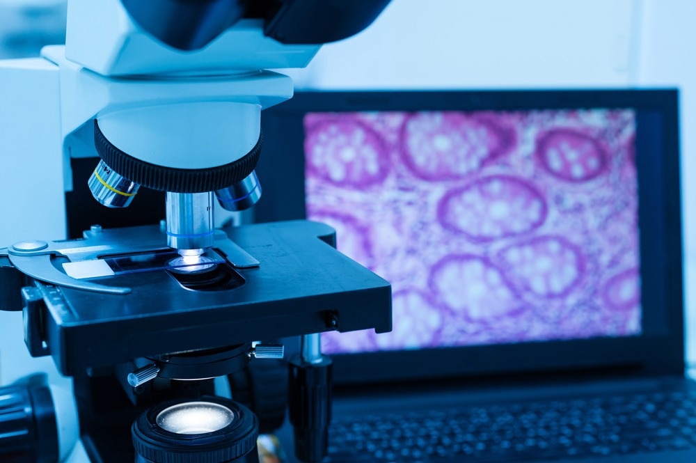
Image Credit:
The birth of digital microscopy
In 1986, the world's first digital microscope was manufactured in Tokyo, Japan. The groundbreaking instrument was built from a control box and lens connected to a camera. This digitization of microscopy added a whole new realm of capabilities to the field. For the first time, a microscope could handle large datasets - the link with the computer opened the door to collecting, storing, and analyzing large data sets. Over the next two decades, scientists worked on improving the digital microscope technology, and in 2005, a more advanced version of the initial digital microscope was created. This updated digital microscope did not require a computer and instead utilized an in-built unit that constituted a screen and computer.
Ten years later, in 2015, scientists launched a new digitalized microscope with an external computer and USB connection, which leveraged the longevity and speed of the external computer. This meant that requirements for external cable connections were cut. In addition, the images produced by this latest update were enhanced by updates to processing software which allowed for adjustments to images such as image brightness, contrast, and scale.
Since its invention, digital microscopy has been benefitting numerous fields of science. The benefits it provides over traditional microscopy include high-resolution images and the production of images in pixels, the production of 2D and 3D image measurements, and high-capacity data storage. In addition, digital microscopes offer a one-stop process of visualizing samples and producing a computer image simultaneously. Finally, in recent years, the cost of digital microscopes has fallen, making the technology more accessible to a wider number of teams.
As a result, scientific fields that rely heavily on microscopes, such as medical and industrial research, have been able to use the technology to further innovation in their sector. Research into cancer, forensics, dental, fetal and embryonic transplant, and microsurgical procedures are some of the key fields that have benefited from the digital evolution of microscopy.
Brands such as Annlov, Palli Partners, Skybasic, and Tomlov, have emerged as industry-leading manufacturers of digital microscopes and are currently relied on by teams of scientists in various research fields.
In the future, we will likely see further developments in digital microscope technology to meet the needs of the research that is reliant upon it. The COVID-19 pandemic, for example, has highlighted the importance of digital microscopy in forensic epidemiology - the study of how diseases are spread. Digital microscopy may continue to evolve to better serve these research areas.
Sources:
- 2019. The Microscope [online]. Science Museum. Available at: www.sciencemuseum.org.uk/objects-and-stories/medicine/microscope (Accessed July 2022)
- Digital Microscopes [online]. Skybasic. Available at: https://skybasic.digitalmicroscopes.us (Accessed July 2022)
- Doherty, M., 2012. Discovering the 'true form:' Hooke's Micrographia and the visual vocabulary of engraved portraits. Notes and Records: the Royal Society Journal of the History of Science, 66(3), pp.211-234. https://royalsocietypublishing.org/doi/10.1098/rsnr.2012.0031
- Microcope products. Annalov. Available at: https://www.annlov.net/c/mircoscope_0031 (Accessed July 2022)
- Shi, W., Georgiou, P., Akram, A., Proute, M., Serhiyenia, T., Kerolos, M., Pradeep, R., Kothur, N. and Khan, S., 2021. Diagnostic Pitfalls of Digital Microscopy Versus Light Microscopy in Gastrointestinal Pathology: A Systematic Review. Cureus,. www.cureus.com/.../65875-diagnostic-pitfalls-of-digital-microscopy-versus-light-microscopy-in-gastrointestinal-pathology-a-systematic-review
- Tomlov [online]. Tomlov. Available at: https://www.tomlov.com (Accessed July 2022)
Further Reading
Last Updated: Nov 21, 2022
 Scientists Track Tiny Human Proteins Using Novel Laser Phase Plate