Polarized light microscopy enhances image contrast and improves image quality in comparison to other microscopy methods, such as differential contrast, phase contrast, and fluorescence microscopy.
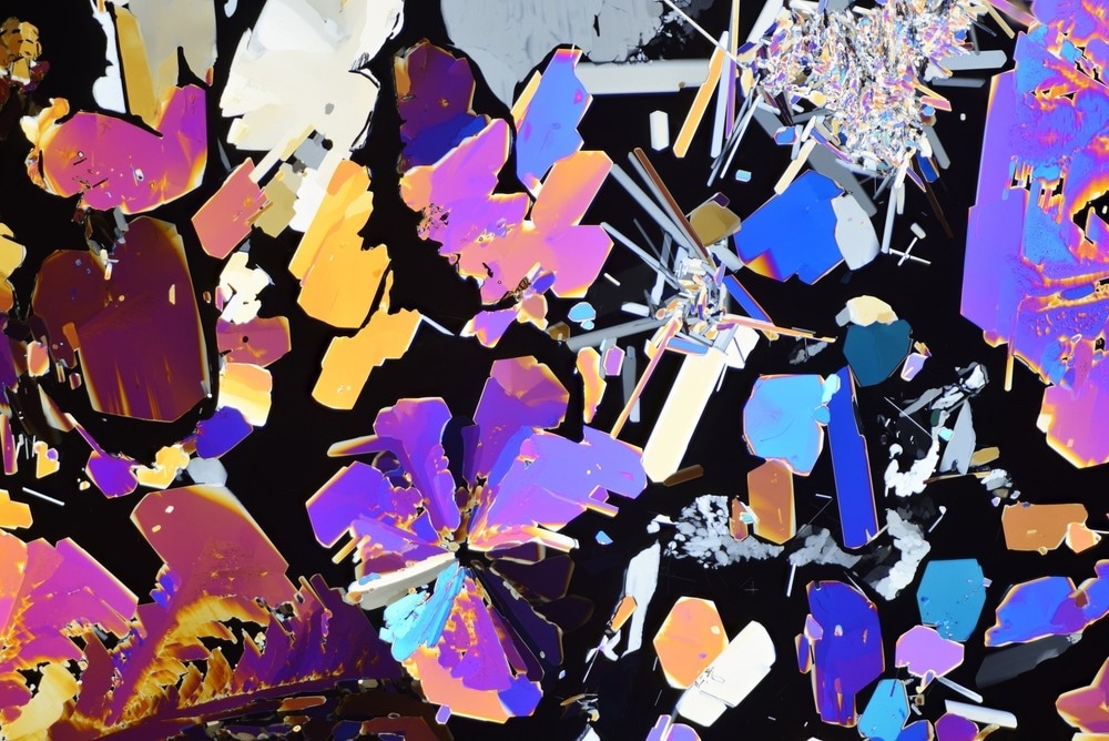
Image Credit: alex7370/Shutterstock.com
What is Polarized Light?
Light is an electromagnetic wave. Although light waves can vibrate in all directions, in general, they are described as vibrating in two directions at right angles to each other.
Any light which vibrates in more than one direction is called ‘unpolarised light’; whereas, a light wave that vibrates in a single direction is called ‘polarised light’. The human eye is not sensitive to the direction of vibration of light.
Converting Non-Polarized to Polarized Light
Polarized light microscopes work by converting unpolarized light to polarized light. One way to achieve this is by absorption of light vibrational movement in one specific direction. Certain natural minerals, including tourmaline, or synthetic films that perform the same function can do this.
Polaroid filters consist of tiny crystallites of iodoquinine sulfate oriented in the same direction and embedded in a polymeric filter. This embedding prevents migration and changes in the orientation of the crystals. The device that selects plane-polarized light from natural or unpolarized light is called a polarizer.
The Principles behind Polarized Light Microscopes
In a polarized light microscope, a polarizer intervenes between the light source and the sample. Thus, the polarized light source is converted into plane-polarized light before it hits the sample. This polarized light falls on a doubly refracting specimen which generates two wave components that are at right angles to each other. These two waves are called ordinary and extraordinary light rays.
The waves pass through the specimen in different phases. They are then combined using constructive and destructive interference, by an analyzer. This leads to the final generation of a high-contrast image.
Components of a Polarized Light Microscope
polarizing microscope
Polarizers
Polarizing filters are the most critical part of the polarized light microscope. There are usually two polarizing filters: the polarizer and the analyzer. The polarizer is located below the specimen stage and can be rotated through 360°. It helps to polarize the light which falls on the specimen.
The analyzer is placed above the objective and may be rotatable in some cases. It combines the different rays emerging from the specimen to generate the final image.
Specialized Stage
This is the specimen stage, and it can rotate 360° to facilitate the specimen's correct orientation with the objective plane. In several stages, a Vernier scale is also provided to provide an accuracy of 0.1° in the stage's rotational angle.
Strain-Free Objectives
Any stress on the objective during installation can lead to a change in the optical properties of the lens which can reduce the performance.
Strain can also be introduced if the lens is mounted too tightly on the frame. Also, anti-reflection coatings and refractive properties must be accurately assessed in order to ensure polarization and increased contrast.
Revolving Nosepiece
As the stage and objectives can revolve in many polarizing microscopes, a revolving nosepiece is also often fitted such that the specimen can be visualized in the center of the view field even if the stage is rotated.
Compensator and Retardation Plates
Several polarization microscopes have compensators and/or retardation plates. This is placed between the crossed polarizers to increase the difference in the optical path in the specimen. This would further increase the contrast of the image quality.
Thus, polarizing microscopes are being used to increase the image contrast to visualize many anisotropic sub-cellular structures.
Conclusion
Polarizing microscopes are widely used to enhance image contrast, making it easier to visualize anisotropic sub-cellular structures.
Further Reading
Last Updated: Oct 3, 2024
 Discovery Challenges the Theory That Proteins Move Mainly by Diffusion