Microscopy plays a vital role in biological research. It optically magnifies minute structures in fixed cells of a tissue. Expansion microscopy (ExM) is an emerging technique that has been developed in the Boyden Laboratory, MIT. One of the key advantages of this microscopy is that it overcomes the limitations associated with optical super-resolution techniques by physically enhancing biological tissues with a water-swellable polymer.
Since the development of ExM, researchers have continually worked on improving the existing range of biomolecules that can be incorporated into the gel network. Additionally, they have focused on the enhancement of the resolving power of ExM.
Expansion Microscopy Explained
A Brief Overview of ExM
ExM technique uniformly expands biological specimens in all dimensions with the help of common reagents and hardware present in most biological laboratories. Put simply; the ExM technique facilitates the isotropic expansion of specimens. The general workflow associated with ExM is as follows:
- Gelling involves the infusion and formation of gel within a biological specimen
- Anchoring of the biomolecules to the gel
- Enzymatic or mechanical homogenization of the specimen
- Physical expansion of the biological specimen.
In most cases, ExM methods employ poly (acrylate-co-acrylamide) gel, which possesses swelling properties when subjected to pure water. Biological specimens are often pre-treated so that biomolecules do not get displaced in the expansion process. In this regard, samples are pre-treated with anchoring reagents that bind biomolecules (e.g., proteins, lipid, and nucleic acids) to the polymer.
Earlier ExM methods involved transferring spatial information of target biomolecules using oligo-conjugated secondary antibodies and reverse-complement trifunctional fluorescent oligos containing a gel-anchorable methacryloyl group. However, current strategies directly link target biomolecules to the gel network via variable bifunctional anchors composed of biomolecule reactive groups and a free-radical reactive group. During polymerization, the free radical reactive group forms a covalent bond with the gel.
Researchers have enabled direct protein retention by using succinimidyl esters (biomolecule reactive group) that interact with primary amines on the surface of protein molecules. As antibodies are proteins, researchers could perform imaging of expanded mouse brain tissue immunostained with commercially available primary antibodies and fluorescent secondary antibodies.
As proteins are directly anchored to the gel, researchers believe that in the absence of proteinase K treatment, protein epitopes could also be preserved for post-expansion immunostaining. The above observation was tested through two different homogenization methods. One method was denaturing and hydrolyzing tissue proteins by incubating the biospecimens in an alkaline detergent solution at 120 °C. The other method used LysC, a type of protease, to conserve epitopes.
A group of researchers has demonstrated a heat denaturation strategy for mechanical homogenization to perform port-expansion labeling. They used a hot detergent solution to homogenize gelled specimens, which better preserved protein epitopes. This technique has been referred to as magnified analysis of the proteome (MAP), which can successfully immunolabel mouse brain tissues after expansion. Besides protein, spatial arrangements of RNA molecules in the brain, at a cellular and subcellular level, play a significant role during normal neural functioning.
The resolution provided by ExM depends on its expansion factor. A higher expansion factor causes higher effective resolution. Scientists adopted two strategies to enhance the expansion factor of a gel. The first strategy is iterative expansion microscopy (iExM), which expands the same biospecimen multiple times. This technique offers around 25 nm effective resolution. Another strategy is X10 microscopy, which requires robust proteinase K digestion for specimen expansion. One of the foremost advantages of ExM is super-resolution imaging deep into a tissue.
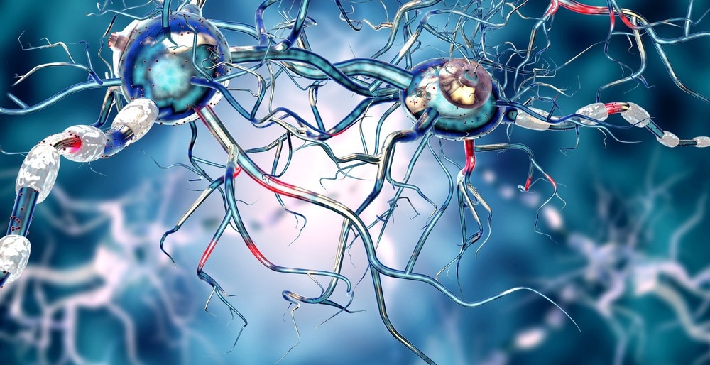
Image Credit: Ralwell/Shutterstock.com
ExM in Neuroscience Applications
Scientists stated that ExM could be applied in a wide range of neurological studies, including transgenic animals. ExM has been applied for imaging synapses and for tracing neurons. Additionally, this technique is used to image a large volume of tissue and neurological diseases.
Identifying putative synapses has been possible; for example, bassoon and homer1 synaptic pair was identified via direct expansion of proteins. To identify the bassoon and homer1 synaptic pair, researchers used fluorophore-conjugated phalloidin and anti-fluorophore antibodies. ExM has been used to perform synaptic imagining of multiple model organisms, including planarian glia, larval zebrafish, and larval and adult Drosophila brains.
The study of local and distal neural connections is extremely important to understand the role of each neuron in circuits during an action. The brain is densely packed with small neural processes, so tracing individual projection to its origin is difficult. Researchers used ExM for successful imaging of a 100-μm column of mouse hippocampal tissue. ExM via the MAP protocol was used to trace and image the whole mouse brain.
Some neurological diseases, such as Parkinson’s and schizophrenia, have been studied using ExM, which is used to determine expanded neurological aberrations or marker proteins.
Future Perspective
Scientists have identified some of the challenges associated with the application of ExM, which have presented opportunities for future research in this area. One of the key bottlenecks of the ExM application is the signal strength in an expanded biospecimen.
Although the expansion of specimens improves the effective resolution, it decreases the concentration of fluorescent tags. Additionally, when the labeling was applied prior to expansion, homogenization, and polymerization, a further loss of fluorophores occurred due to oxidation and hydrolysis of antibodies. Hence, in the future, researchers should focus on improving the resolution offered by ExM. A growing need for imaging the cleared/expanded brain raises the requirement to develop optical objectives, particularly to image large samples.
Sources:
- Gallaggher, R.B. and Zhao, Y. (2021) Expansion microscopy: A powerful nanoscale imaging tool for neuroscientists. Neurobiology of Disease,154. https://doi.org/10.1016/j.nbd.2021.105362
- Kubalová, I. et al. (2020) Prospects and limitations of expansion microscopy in chromatin ultrastructure determination. Chromosome Research, 28, pp. 355–368. https://doi.org/10.1007/s10577-020-09637-y
- Götz, R. et al. (2020) Expansion Microscopy for Cell Biology Analysis in Fungi. Frontiers in Microbiology. 11. DOI=10.3389/fmicb.2020.00574
- Wassie, A.T. et al. (2019) Expansion microscopy: principles and uses in biological research. Nature Methods, 16, pp. 33–41. https://doi.org/10.1038/s41592-018-0219-4
- Klimas, A. et al. (2019). Basics of Expansion Microscopy. Current protocols in cytometry, 91(1). e67. https://doi.org/10.1002/cpcy.67
- Chen, F. et al. (2015) Expansion microscopy. Science, 347(6221), pp. 543-548.DOI: 10.1126/science.1260088
Further Reading
Last Updated: Jul 12, 2022
 Scientists Decode How T Cells Stop Deadly Viruses