A feature of modern life is a desire for smaller and smaller devices to be more and more powerful. Technology can now operate at a tiny nanoscale. There is also a thirst for knowledge about the composition and structure of materials whilst being able to understand them.
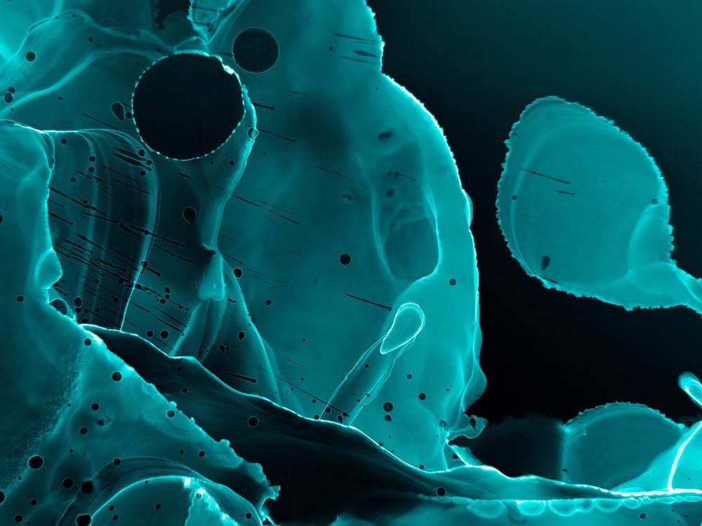
Image Credit: Holo Art/Shutterstock.com
We want to be able to see these tiny things. This desire has led to the development of many forms of microscopy such as optical microscopy, scanning electron microscopy, transmission electron microscopy, scanning probe microscopy, scanning acoustic microscopy, and X-ray microscopy. All of these have pros and cons and are best for particular studies. Often studies will use a combination of microscopes to achieve their goals.
This article will overview one specialist type of microscope, the X-ray microscope. This type of microscope typically operates between optical and electron microscopes in terms of sensitivity, and X-ray microscopes have a resolution down to around 30 nanometers.
Rontgen discovered X-rays in 1895, and Sir Lawrence Bragg produced the first usable X-ray image in the late 1940s. In the 1950s, Sterling Newberry made the first shadow X-ray microscope. The first use of a synchrotron in a microscope was in 1972. However, in the 1990s, improved technology led to the rapid development of the x-ray microscope. The General Electric Company first commercialized x-ray microscopes in the 1950s, but now many companies such as Zeiss, Horiba, and Bruker manufacture X-ray microscopes.
X-Ray microscopes have greater magnification than optical microscopes but lower magnification than electron microscopes. Although the magnification is not as great as electron microscopes, X-ray microscopes have some advantages over them. X-ray microscopes can examine wet samples and, therefore, can be used to study plants, insects, and human cells. They can also be used on slightly thicker specimens and utilize diffraction, refraction, and reflection to obtain images. They can produce 2D or 3D images. However, X-ray radiation can damage cells, so caution must be exercised when examining biological cells. X-ray microscopes also operate at atmospheric pressure and don’t require a vacuum that some electron microscopes do.
Samples in X-ray microscopy do not need as much special treatment as that required by Transmission Electron Microscopes. Samples can be more than the one-micron thickness limit of Transmission Electron Microscopy. It allows for non-destructive visualization of samples, combined with tomography to produce 3-dimensional images of the examined specimen.
X-ray microscopes work by exposing a sample to X-rays. The X-rays will either pass through the sample or to some degree will be absorbed. X-rays that pass through the specimen can then expose a film or charge detector device to produce a visible image. Techniques like absorption spectromicroscopy, microspectroscopy, and others generate detailed images in modern x-ray microscopes. They generally run at ambient temperature and pressure and use a standard 220 volt supply amplified by a transformer to many kilovolts to produce x-rays. X-ray microscopes may be whole field or Scanning X-Ray Microscopes.
Types of X-Ray Microscope
There are two main types of X-ray microscope;
Submicron X-ray microscopes use a two-stage magnification. An X-ray beam is projected onto a sample and produces an image projected behind the sample and captured on film or an electronic device. It is an x-ray shadow projection model which will give images down to 700-nanometer resolution.
Nanoscale x ray microscopes. In this type of microscope, the X-rays are focused after the sample using Fresnel zone plates to diffract the X-ray beams creating images down to 50 nanometers.
Uses of X-Ray Microscopes
X-ray microscopy is used in materials science to examine the microstructure of porous electrodes in battery and fuel cells both in research and in manufacturing quality control. It is also used to study materials under stress to help understand how they react to impact, bending, or torsion. The analysis of the structures of polymers like polyurethane also relies on X-ray microscopes.
X-ray microscopy can be useful in forensic science to identify tiny fragments, gunshot residue, paint fragments, and elemental makeup of samples in a non-destructive way. The ability to view inside small particles can be significant.
In environmental science, X-ray microscopy can help study biofilms, which are an important tool in bioremediation. The distribution of macromolecular compounds (proteins, carbohydrates, lipids, and nucleic acids) within the cells and the extra-cellular matrix are identified using X-ray microscopy. Biofilms are a typical habitat for microbes in many biological systems and can cause mechanical and corrosion problems and harbor pathogenic organisms in engineered water systems. Identifying their components can help control them.
X-ray microscopy can be used to view sub-cellular structures and is used to view chromosomes and other small biological bodies.
Soil structure is studied using X-ray microscopy to determine the structures of the soil. It is used in geochemistry, cosmochemistry, polymer science, and biology to determine structures. Magnetism and materials science also use X-ray microscopy for various reasons.
X-ray microscopes are an important tool in many sciences and industries allowing nondestructive testing of small samples. Whilst they don’t have the magnification of an electron microscope they are easier to use in many respects and are a vital tool in many situations.
Further Reading
Last Updated: May 5, 2022
 Real-Time Molecular Tool Observes Complex Immune System Changes