Reviewed by Lexie CornerMay 14 2025
Researchers at the Terasaki Institute for Biomedical Innovation (TIBI) have developed a technique that may improve tissue engineering approaches. Their findings outline a method for generating tissues with controlled cellular organization, aiming to replicate the natural structure of human tissue.

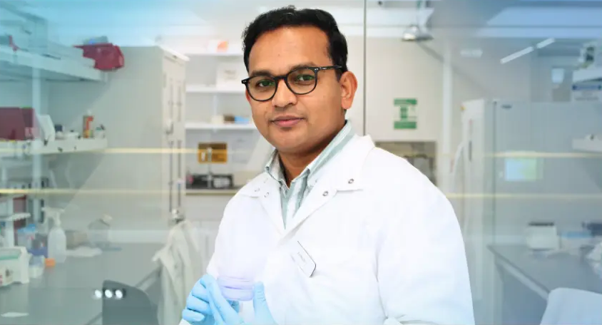
Image Credit: Terasaki Institute for Biomedical Innovation
Using a light-based 3D printing method, the team fabricated microgels with controlled internal structures that influenced cell behavior and growth, mimicking the natural dynamics of cells in the body.
By adjusting how light interacts with hydrogels, they were able to modify the internal architecture of the microgels, enabling precise control over cell organization in three dimensions.
This approach addresses a key challenge in creating realistic, functional tissue environments needed for effective tissue repair and regeneration.
Our technique enables the production of microtissue with precise structural control, which is essential for engineering tissues such as muscle and retina. We are enabling a new class of modular biomaterials that can actively guide tissue formation and engineering organ through the bottom-up approach.
Dr. Johnson John, Study Principal Investigator, Terasaki Institute for Biomedical Innovation
The study showed that these microgels could be adapted for a range of biomedical applications. In one example, muscle cells embedded within rod-shaped gels aligned and formed muscle fibers, marking a promising step toward injectable therapies for muscle injuries.
In another case, the gels were used to encapsulate photoreceptor cells, which self-organized into layered structures resembling the outer retina, highlighting the potential for future retinal treatments.
The team also incorporated angiogenic peptides into the gels, encouraging new blood vessel formation both in vitro and in vivo. The microgels maintain their shape during injection and are designed to support cell growth, vascularization, and tissue formation.
Their flexible design allows for tailoring to specific medical needs, making them a promising platform for wound healing, organ repair, and disease modeling.
This work represents a significant step toward creating structures that can form functional tissues. By merging light-based fabrication with smart biomaterials, we are getting closer to making personalized, minimally invasive therapies.
Dr. Ali Khademhosseini, Chief Executive Officer, Terasaki Institute for Biomedical Innovation