Researchers at ETH Zurich have achieved an unprecedented live, high-resolution visualization of influenza virus infection within living cells. This milestone was made possible by an innovative microscopy technique, which is expected to facilitate the more targeted development of antiviral therapies.
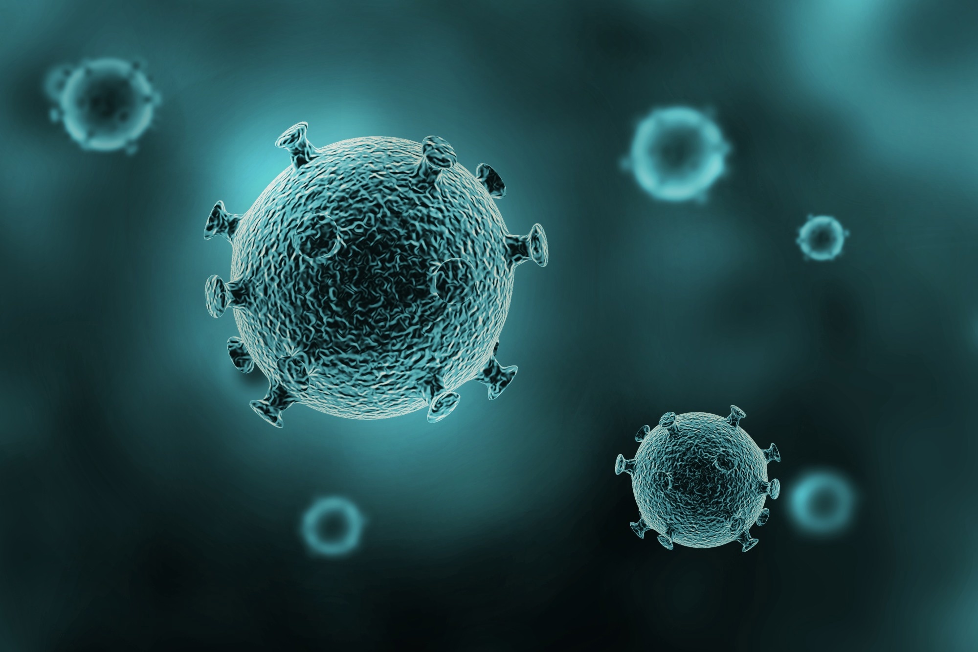 Image credit: sumroeng chinnapan/Shutterstock.com
Flu, characterized by symptoms such as fever, myalgia, and rhinorrhea, recurs seasonally. This disease is caused by influenza viruses, which are transmitted via respiratory droplets and subsequently infect host cells.
A collaborative research effort by Swiss and Japanese scientists has examined the influenza virus. Employing a novel, self-developed microscopy technique, these researchers achieved high-resolution, live observation of influenza virus entry into human cells cultured in a Petri dish, marking a significant advancement in real-time cellular imaging.
Under the direction of Yohei Yamauchi, Professor of Molecular Medicine at ETH Zurich, a notable discovery emerged: host cells do not passively permit influenza virus invasion. Instead, they actively engage in the capture of the virus.
The infection of our body cells is like a dance between virus and cell.
Yohei Yamauchi, Professor, Molecular Medicine, ETH Zurich
 Cells actively help to capture and incorporate influenza viruses. Here, a cell is shown, with a virus in the center of the image. Image Credit: Emma Hyde / ETH Zurich
Viruses Surf on the Cell Surface
Cellular infection by viruses does not confer any advantage to the host cell, nor does the cell actively participate in the infection process. Instead, viruses exploit an essential, everyday cellular uptake mechanism. This critical mechanism is typically responsible for internalizing vital substances, including hormones, cholesterol, and iron, into the cell.
Similar to these vital substances, influenza viruses initiate infection by attaching to specific molecules on the cell surface. This interaction involves the virus systematically scanning the surface, transiently binding to various molecules until an optimal entry point is identified. Such a point is characterized by a high localized concentration of receptor molecules, facilitating efficient cellular uptake.
Upon detection of viral attachment by cellular receptors on the membrane, a localized depression or pocket begins to form. This invagination is structurally shaped and stabilized by the specialized protein clathrin. As this pocket deepens, it fully encloses the virus, culminating in the formation of a vesicle. The cell subsequently internalizes this vesicle, and once inside, the vesicle coating disassembles, releasing the virus into the cellular interior.
Investigations into this critical cellular process have historically relied on microscopy techniques such as electron microscopy. These methods necessitate cell destruction, thereby yielding only static snapshots of the dynamic event. Fluorescence microscopy, another employed technique, is limited by its inherently low spatial resolution.
Combined Techniques, Including for Other Viruses
A novel technique, virus-view dual confocal and AFM (ViViD-AFM), integrates atomic force microscopy (AFM) and fluorescence microscopy. This method now enables the detailed observation of viral entry dynamics into cells.
Research findings demonstrate that the cell actively promotes multi-level virus uptake. This process involves the active recruitment of clathrin proteins to the virus's location. The cell surface actively captures the virus through localized bulging. These wavelike membrane movements intensify when the virus attempts to detach from the cell surface.
This novel technique offers critical insights for antiviral drug development. Its utility extends to real-time efficacy testing of prospective drugs within cell cultures. The study's authors highlight the technique's potential application in investigating the behavior of other viruses and vaccines.
How influenza viruses enter our cells
Video Credit: Nicole Davidson / ETH Zurich
Source:
Journal reference:
Yoshida, A., et al. (2025) Enhanced visualization of influenza a virus entry into living cells using virus-view atomic force microscopy. PNAS. DOI:10.1073/pnas.2500660122. https://www.pnas.org/doi/10.1073/pnas.2500660122.
 Microtubule-Stabilizing Protein Camsap3 Proves Essential for Female Fertility