In this interview, AZoLife Sciences speaks with Elodie Litzler, Co-Founder and Chief Operating Officer at Avatar Medical, about how advanced 3D visualization is transforming medical imaging into an intuitive tool for clinicians and patients alike.
Could you start by introducing yourself and telling us what Avatar Medical is all about?
I’m Elodie Litzler, Co-Founder and COO of Avatar Medical. The company was a spin-off from Institut Pasteur and Institut Curie in Paris. Our team started with a simple question: what if every CT or MRI scan could be turned into a clear, intuitive 3D view that both non-radiologist physicians and patients can instantly access and understand?
Avatar Medical turns routine DICOM data into clear, interactive 3D “avatars” that make anatomy instantly understandable, even when no radiology expertise is available, whether you’re a surgeon preparing a complex case or a patient trying to make sense of a diagnosis. Our work is about bridging the long-standing gap between what is shown in imaging and what is truly understood, and providing every clinician and patient with a shared, intuitive view of what’s happening inside the body.
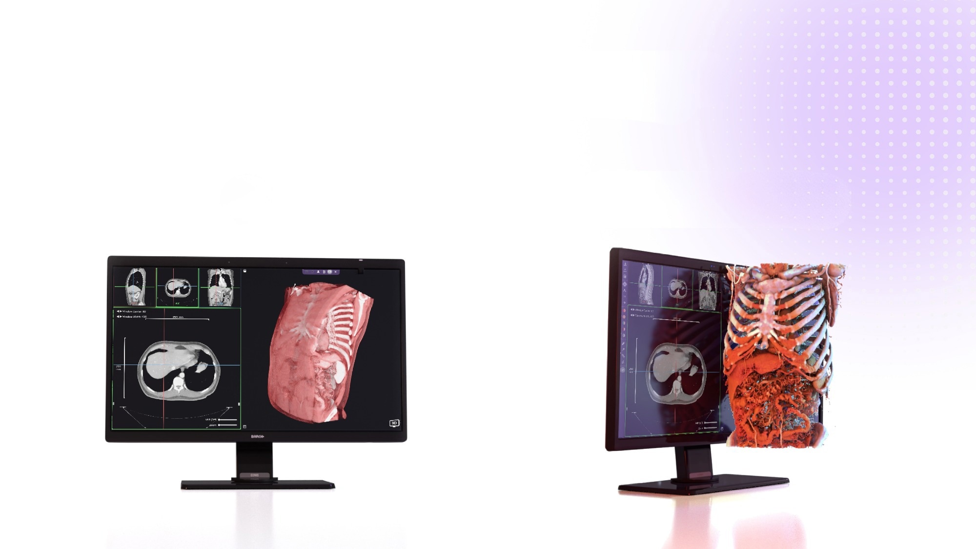 Image credit: Avatar Medical
What gap did you see in medical imaging or patient care that motivated you to found Avatar Medical in 2020?
CTs and MRIs offer extraordinary information, but the way that information is typically shared, 2D grayscale slices, hasn’t evolved much in decades. While radiologists are trained to interpret these, other physicians, especially patients, often struggle. Even when 3D imaging tools exist, they’re usually made for radiologists, require training, or rely on flat screens that lack depth perception. This can be challenging to understand complex pathological anatomies, like a blood vessel wrapping a tumor.
However, some clinicians really benefit from 3D representations, such as surgeons who must interact in real-world space with 3D patients. However, this imaging is not yet available everywhere. The tools and reimbursement, when they exist, are mostly in radiology. For a radiologist, creating a 3D model for a surgeon requires time, often does not pay well, or does not pay at all, and there is a global shortage of radiologists.
The issue isn't a lack of 3D technology, but a lack of access and usability.
We recognized the challenges of mentally reconstructing 3D anatomy from flat images (see our publication in JCO for Breast surgeons). This lack of spatial cognition is especially concerning because surgical approaches can be based on such interpretations, and some exploratory surgeries or surgical mistakes are made every day due to this. Not to mention the bandwidth required to create a model for individual consultations as a visual aid for patients and informed decision-making.
Our interactions with dozens of surgeons led us to believe that having a stereoscopic display, which displays one slightly different image for each eye, allowing the brain to reconstruct a perception with depth, would improve their understanding, even if no clinical benefits had been proven at the time.
It just felt obvious: if you have 3D data, why observe it through slices or flat 3D projections on a flat screen, when you could see it at scale with stereoscopy? This motivated us to create Avatar.
Read more about how Avatar Medical is transforming medical imaging. Download your PDF copy now!
What’s a misconception about medical imaging or 3D visualization that clinicians or people hold?
Among clinicians, many still believe that 3D imaging requires expensive equipment, lengthy processing times, and specialized training. That was historically true, but recent advances from industries like gaming have changed that.
With today’s graphic processing units (GPUs) and stereoscopic 3D rendering technologies, high-quality 3D visualization can now be fast, interactive, and affordable.
As for patients, most are simply unaware that their own scans can be turned into personalized 3D models. They're used to grayscale slices and don’t realize you can make a personalized 3D model out of their data.
For people who may not know your work yet, how would you describe Avatar Medical and what your team is trying to achieve?
We turn standard CT or MRI DICOM files into interactive, highly accurate 3D anatomical models in minutes. Our goal is to bring clarity to every point of care, whether that’s surgical planning, patient education, or multidisciplinary discussions.
By reducing cognitive load for clinicians and making medical imaging understandable for patients, we enable better communication, better preparation, and ultimately better outcomes.
Could you walk us through the technical pipeline of Eonis Vision - from CT/MRI DICOM data to the final glasses-free 3D anatomical “avatar.”
Eonis Vision combines three key components to create a seamless experience: Avatar Medical’s 3D visualization engine, Barco’s glasses-free 3D display, and the consistent compute performance provided by Dell. Here’s how it works:
Import
The clinician uploads standard CT or MRI DICOM files; no special imaging acquisition settings or new workflows are required.
Processing
Our algorithms automatically generate a 3D model from the imaging data in seconds. This is where reliable compute power matters: the system needs to process large imaging datasets quickly and consistently. Dell provides the behind-the-scenes performance that allows this to happen without delay or complexity.
Optimization for 3D
In one click, the doctor selects an anatomical region of interest in the grayscale image, and an adapted filter is applied to the 3D representation so the model is visually ready for clinical exploration and discussion. This is our key intellectual property. Our user experience is similar to using a slider in your smartphone photo app. Even if you don’t know what the algorithm is doing in the background, you can stop touching the slider when you are happy with the representation. No need for any intense training to do that.
Display on Eonis Vision
Barco’s glasses-free 3D display enables the final avatar to appear with natural depth perception, eliminating the need for headsets or glasses. Physicians and patients simply step up to the display and immediately see the anatomy in true 3D.
It’s a seamless experience, where each partner contributes key technology to make medical imaging truly accessible. Avatar Medical transforms the data, Dell ensures the experience is smooth and reliable, and Barco brings it to life in a way that feels intuitive in any exam room.
How does the collaboration with Barco improve what you can offer?
From the start, we tested all available stereoscopic technologies, including virtual reality (VR) headsets, augmented reality (AR) glasses, and 3D displays; however, they all required the user to wear a device. That’s not practical in clinical consultations or with patients.
They all had the same limitation: you need to wear a device to get stereoscopy. This was making it difficult to implement in consultations. It takes a long time, requiring disinfection between patients. Another limitation is that they are consumer electronics devices and not stable medical devices.
Barco’s Eonis Vision changes everything. It’s a medical-grade, glasses-free 3D display that switches between 2D and 3D at the click of a button. This switchability is enabled by Leia technology, which is embedded in Eonis 3D. This makes it easier to integrate into consultations and use with patients. Combined with Dell’s stable compute infrastructure, we have a complete system that’s practical, scalable, and intuitive. It is also a clinical-grade device, more suitable for the medical environment.
What feedback have you received from surgeons, radiologists, or patients using Avatar Medical’s 3D visualizations? Any striking success stories or surprising use cases
What we hear most from surgeons is that the technology changes how they prepare and how their patients understand what’s ahead of them. But the most powerful feedback always comes from patients themselves.
Patients today come to consultations with higher expectations. There is a wealth of information available to patients today, which can be overwhelming and confusing to navigate.
We’ve had people cry from relief after finally understanding their condition through 3D visualization. They no longer feel lost in technical jargon. They become active participants in their care. This emotional clarity, this ability to truly see what’s happening inside their own body, is why we created Avatar Medical.
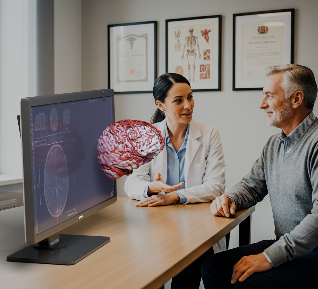
Image credit: Avatar Medical
How do you ensure accessibility and equity ( i.e. making sure this technology benefits not only top-tier hospitals but also smaller clinics, community hospitals, or resource-limited settings?)
The latest medical technology is often first introduced in top-tier hospitals. This is, unfortunately, the standard approach.
However, as adoption grows and technology develops, costs come down, just as with surgical robots or VR headsets.
In the case of Eonis Vision, it can be expected that the cost of the hardware will continue to decrease, as it has for GPUs and VR headsets. The cost of the software licences can also go down as the global volume increases and the supporting digital service infrastructure develops.
Beyond oncology and surgical planning, do you see potential for Avatar Medical’s technology in other domains
The core technology is generic and can be applied to any CT and MRI.
We started exploring neurosurgery and ENT oncologic surgery in the US. However, I am convinced it will have an impact anywhere where 3D imaging is the standard for diagnostics, which is becoming the case as the global number of machines increases. In Europe, we have it deployed for breast cancer surgery consultations at the American Hospital of Paris.
We’ve recently begun pilots in orthopedic consultations as well. The need for understanding is universal, no matter the specialty, patients want clarity before undergoing surgery or treatment.
Are there any next-generation features or functionalities you’re working on?
Yes, we’re working on controller-free manipulation using technology from other industries, such as hand tracking, letting users rotate and zoom the models without touching a device. This is especially useful in sterile environments like operating rooms.
And AI, of course. The improvements in medical image analytics through AI have been incredible over the past ten years. We will not invent new AI techniques, but we will work on making the results of AI algorithms available in our products, which means displaying annotations, segmentations, and measurements at the top of our lossless renderings. An interesting way for physicians to monitor the accuracy of AI.
Download your PDF copy now!
Further Reading
To learn more about Avatar Medical, visit here.
To see and explore how Eonis Vision is the next generation of AR for the clinic, visit here.
For Avatar Medical's insights on patient experience advanced visualization, visit here.
About the Researcher:

Elodie Litzler has built her career at the intersection of research, operations, and innovation. At the Institut Pasteur, she led operations as Deputy CTO, spearheaded the adoption of cutting-edge technologies, and later transitioned into the Innovation Office to help turn academic research into viable prototypes. Her work on the DIVA project ultimately supported the creation of Avatar Medical in 2020. Since then, she leads the Engineering, Product, and QARA teams; secured over €4M in grants, earned the company’s first FDA clearance, and established clinical partnerships across France.
 Creatine Linked to Cancer-Fighting Dendritic Cell Function