Key Components, Principles, and Challenges of Early Bioprinting
The Road to Real-World Bioprinting
Bioinks formulation
Printing technology
Smarter biomaterials
Challenges in Clinical Adoption of Bioprinted Tissues
References and Further Reading
Researchers are overcoming long-standing barriers in tissue engineering by developing bioinks and smart hydrogels that actively support vascular growth, cellular maturation, and tissue stability after printing. These advances are positioning bioprinting as a scalable platform for personalized regenerative therapies, advanced disease models, and future organ fabrication.
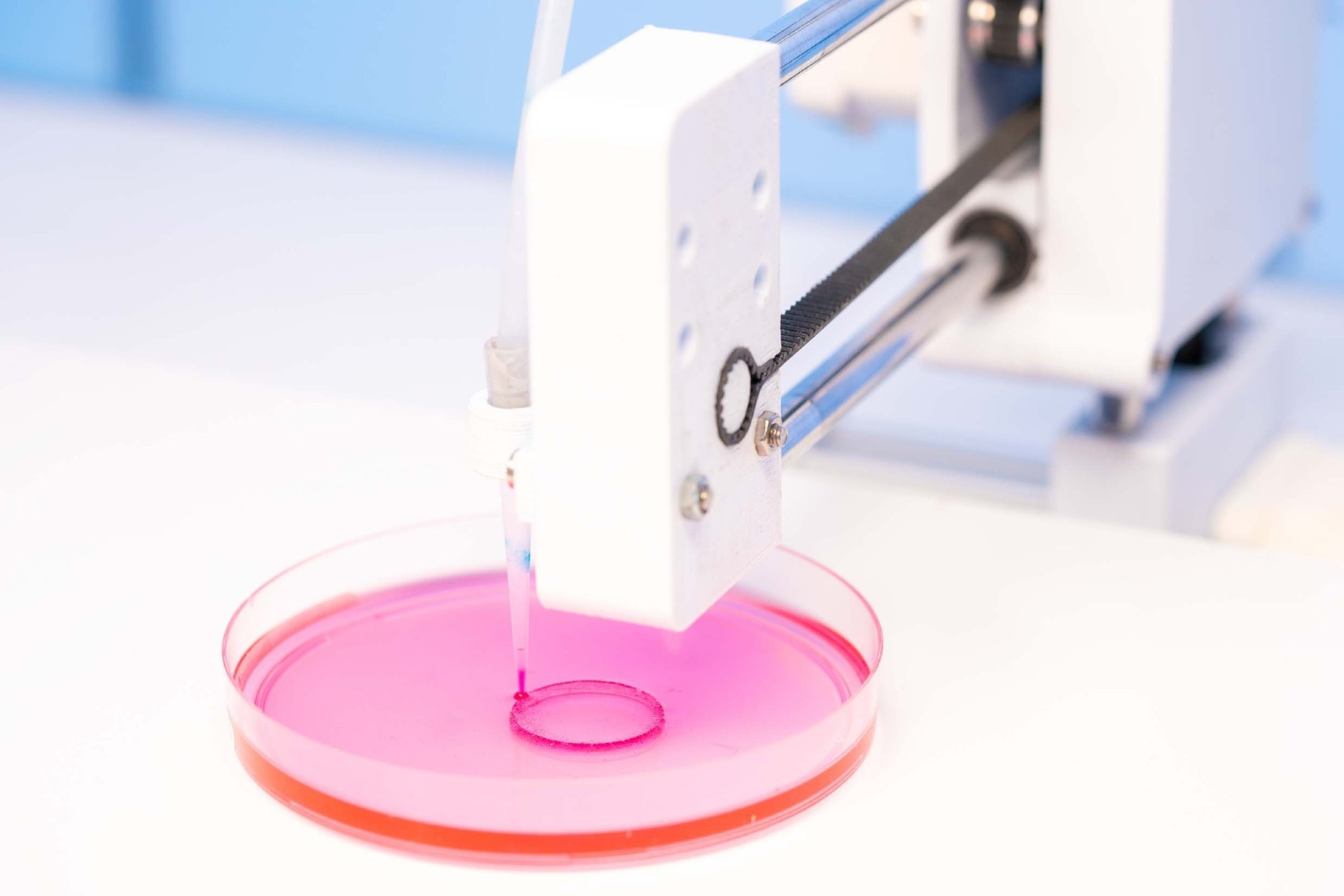 Image credit: luchschenF/Shutterstock.com
Bioprinting has evolved into a leading biomedical technology by combining advanced bioink formulations, innovative printing methods, and smart biomaterials. These breakthroughs have enabled the rapid creation of complex, functional tissue constructs that bring regenerative medicine and personalized therapies closer to clinical reality.
Key Components, Principles, and Challenges of Early Bioprinting
Bioprinting is an additive manufacturing technique that fabricates living tissues by assembling cells and biomaterials, layer by layer or volumetrically, to mimic native biological structures. Bioprinting is formally defined as the use of material transfer processes to pattern and assemble biologically relevant materials, such as living cells and biomolecules, into organized functional structures. Key components of bioprinting include bioinks, which combine living cells and supportive biomaterials designed for tissue growth, and the cells selected for the target tissue.1,3
Bioprinter hardware uses digital models, such as Computer-Aided Design (CAD) files or 3D models, to represent the precise geometry of the biological structure or tissue to be printed. Specialized software is used to make these models and break them into thin layers. The bioprinter then follows these layers to place materials precisely, enabling it to build complex structures such as tissues, organs, and scaffolds according to a digital plan. Structural support, typically provided by scaffolds, helps maintain the shape of the printed constructs. After printing, crosslinking methods are used to stabilize the tissue, and bioreactors or culture systems aid its maturation and function.
Historically, bioprinting has shown promise for regenerative medicine, but progress has been limited by unsuitable biomaterials and the technical complexity of creating viable tissue structures. Early efforts produced only simple constructs due to inadequate bioinks and slow, imprecise printing methods that struggled to mimic native organs. Traditional tissue engineering methods also faced challenges, including uneven cell distribution, low cell density, and limited integration of vascular and neural networks.2
Find out more about the key concepts of bioprinting. Download your free PDF article here
The Road to Real-World Bioprinting
Recent advancements in bioink formulation, printing technology, and biomaterials have propelled bioprinting beyond its experimental origins toward practical applications in medicine. This progress is enabling breakthroughs in tissue repair, organ transplantation, disease modeling, and drug testing. Key advances facilitating real-world bioprinting are discussed below:
Traditional bioinks such as alginate, agarose, and certain synthetic hydrogels often lack the strength, cell compatibility, and tissue-mimicking properties required for optimal cell survival and print quality. Recent breakthroughs in bioink formulation have created materials that better mimic the natural cellular environment, support cell viability and differentiation, and enable the construction of more complex tissues.2
Hydrogels derived from natural polymers such as collagen, hyaluronic acid, alginate, and gelatin are especially important because they provide hydrated extracellular matrix-like environments that support cell attachment, proliferation, migration, and differentiation. These biomaterials can also be supplemented with growth factors, bioactive molecules, and multiple cell populations to better replicate the complexity of native tissues.1,3
Protein-based materials have emerged as leading candidates for bioink formulation, offering a wide range of tunable properties. Natural proteins such as silk fibroin, elastin, resilin, collagen, and gelatin stand out for their intrinsic mechanical strength and elasticity, making them especially valuable for 3D printing applications. Protein-based bioinks have enabled innovations such as magnetically responsive hydrogels, in situ bioprinting, and 4D bioprinting, which can replicate the structure and dynamic behavior of real tissues. Ongoing progress in developing multicomponent, protein-rich bioinks is expected to further enhance print fidelity, tissue integration, and clinical applicability in advanced tissue engineering.3
A major focus of current bioink research is recreating the extracellular matrix (ECM), a dynamic biological network composed of collagens, proteoglycans, elastin, and glycoproteins that regulates cellular adhesion, signaling, migration, and tissue organization.3
Modern bioink systems are increasingly designed to incorporate biochemical and mechanical cues that regulate cell fate, including stiffness, degradability, nutrient diffusion, and shear-thinning behavior during extrusion. These properties improve printability while minimizing mechanical stress on encapsulated cells and preserving cell viability.8
Printing technology
Traditional bioprinting techniques, such as extrusion and inkjet printing, often face significant limitations. These methods commonly involve layer-by-layer deposition, which can result in slow fabrication speeds, limited structural complexity, and challenges in maintaining cell viability due to mechanical stress and prolonged processing times. Additionally, the resolution and fidelity of complex 3D structures are limited by nozzle size and printing path, making it difficult to reproduce intricate tissue architectures found in nature.4
Volumetric bioprinting is a rapidly evolving technique that uses light to create 3D structures from soft, cell-containing hydrogels. This approach enables fast and precise construction of complex tissues for medical research and regenerative medicine. Unlike conventional layer-by-layer approaches, volumetric bioprinting can fabricate centimeter-scale constructs within seconds by using tomographic light projections to selectively crosslink photoresponsive bioresins. This rapid fabrication process reduces mechanical stress on fragile cells and improves the production of geometrically complex tissue structures.4,5
Recent advances have enabled multi-material printing, which is important for building realistic tissue models. One particularly significant direction is the combination of volumetric bioprinting with extrusion printing in supportive baths. This hybrid approach leverages the speed and complexity of volumetric methods with the spatial control and material versatility of extrusion, enabling the fabrication of more intricate and heterogeneous tissue constructs than previously possible. Emerging embedded extrusion volumetric printing approaches also enable the simultaneous positioning of multiple cell populations and biomaterials within supportive hydrogel baths, improving the fabrication of heterocellular and anatomically complex tissues.4,6
Current bioprinting workflows frequently integrate advanced imaging technologies such as MRI, CT, and X-ray scanning to generate patient-specific digital models that guide precise tissue fabrication.8
How do you print 3D organs and tissue? Watch this.
Video credit: ASME American Society of Mechanical Engineers/Youtube.com
Smarter biomaterials
Traditional biomaterials used in bioprinting, including natural and synthetic polymers, have long been valued for their biocompatibility and ease of processing. However, a fundamental limitation of these materials is their static nature. They are unable to actively respond or adapt to changes in their environment, which restricts their utility in applications that demand real-time responsiveness or structural adaptability, such as regenerative medicine or tissue engineering.7
In contrast, next-generation smart hydrogels represent a significant leap forward. These materials are engineered to sense and respond to environmental stimuli, such as changes in pH, temperature, or glucose concentration, enabling tailored, precise functionality. For instance, glucose-responsive hydrogels are being developed for diabetic wound care, where they alter drug release in response to local glucose fluctuations to enhance therapeutic outcomes.8
Recent glucose-responsive hydrogel systems have demonstrated the ability to release insulin adaptively within hyperglycemic wound environments, promoting angiogenesis, cell proliferation, and accelerated diabetic wound healing. Some dual-network hydrogels use dynamic phenylboronic ester bonds together with calcium-mediated crosslinking to improve both glucose sensitivity and mechanical stability.8
Similarly, self-healing hydrogels can autonomously repair structural damage, thereby maintaining material integrity under mechanical stress or injury. Self-healing hydrogels rely on reversible dynamic covalent bonds and noncovalent interactions that allow damaged structures to autonomously restore their integrity and functionality. These biomaterials can also be engineered as injectable, stimulus-responsive, drug-loaded, or shear-thinning systems for applications in tissue engineering and controlled therapeutic delivery.9
Advanced hydrogel systems can also be engineered with tunable stiffness, degradation rates, and biochemical signaling properties that actively guide vascular maturation, tissue remodeling, and long-term integration.6
The advent of these adaptive and intelligent hydrogels is transforming the landscape of biomedical applications by offering new possibilities for personalized therapies, advanced wound dressings, and dynamic tissue scaffolds.
Challenges in Clinical Adoption of Bioprinted Tissues
The primary bottleneck for bringing bioprinted tissues to clinical use is achieving effective vascularization. While bioprinting enables precise architectural assembly of vascular networks, most current methods prioritize geometric accuracy over reproducing the complex, dynamic biological processes required for truly functional blood vessels.10
Hydrogels, designed as active microenvironments, are engineered to deliver the biochemical and mechanical cues necessary for vascular maturation, yet crucial events like lumen formation and cellular remodeling are often missed due to insufficient time-resolved evaluation.10
Functional vascularization requires more than perfusable channels; vessels must mature, remodel, recruit supporting mural cells such as pericytes and smooth muscle cells, and integrate with host tissue over time. Tissue-specific vascular structures also differ substantially, as brain vasculature, liver sinusoids, and pancreatic capillaries each exhibit distinct permeability and structural requirements.10
Transitioning bioprinted tissues from lab to clinic also presents significant translational challenges. Many constructs fall under the category of combination products, incorporating both living cells and biomaterials, which adds complexity to regulatory approval processes.11
Demonstrating safety, reproducibility, and durability of function is challenging, especially as agencies such as the U.S. Food and Drug Administration (FDA) and the European Medicines Agency (EMA) adapt their frameworks for these novel products. Regulatory agencies also face challenges related to classification, quality control, manufacturing standardization, software validation, and oversight of personalized patient-specific constructs. Because bioprinted products combine the complexities of additive manufacturing, biomaterials, stem cell technologies, and regenerative medicine, existing regulatory systems are often poorly suited to their evaluation.11
Additional barriers include manufacturing scalability, cost-effectiveness, ethical considerations, and the long-term maintenance of viable multicellular tissue structures. Ultimately, clinical adoption will depend on successfully uniting biomaterial-driven biological cues with high-precision bioprinting and meeting evolving regulatory standards.4
References and Further Reading
- Tripathi S, et al. 3D bioprinting and its innovative approach for biomedical applications. MedComm. 2022;4(1):e194. DOI:10.1002/mco2.194, https://onlinelibrary.wiley.com/doi/10.1002/mco2.194.
- Murenu N, et al. Bioprinting Organs-Science or Fiction? A Review From Students to Students. Adv Healthc Mater. 2026;15(16):e02103. DOI:10.1002/adhm.202502103, https://advanced.onlinelibrary.wiley.com/doi/10.1002/adhm.202502103.
- Veiga A, et al. Current Trends on Protein Driven Bioinks for 3D Printing. Pharmaceutics. 2021;13(9):1444. DOI:10.3390/pharmaceutics13091444, https://www.mdpi.com/1999-4923/13/9/1444.
- Veeravalli RS, et al. Three-Dimensional Bioprinting in Medicine: A Comprehensive Overview of Current Progress and Challenges Faced. Cureus. 2023;15(7):e41624. DOI:10.7759/cureus.41624, https://www.cureus.com/articles/167067-three-dimensional-bioprinting-in-medicine-a-comprehensive-overview-of-current-progress-and-challenges-faced.
- Jing S, et al. Advances in volumetric bioprinting. Biofabrication. 2023;16(1). DOI:10.1088/1758-5090/ad0978, https://iopscience.iop.org/article/10.1088/1758-5090/ad0978.
- Ribezzi D, et al. Multi-material Volumetric Bioprinting and Plug-and-play Suspension Bath Biofabrication via Bioresin Molecular Weight Tuning and via Multiwavelength Alignment Optics. Adv Mater. 2025;37(13):e2409355. DOI:10.1002/adma.202409355, https://advanced.onlinelibrary.wiley.com/doi/10.1002/adma.202409355.
- Chen XB, et al. Biomaterials / bioinks and extrusion bioprinting. Bioact Mater. 2023;28:511-536. DOI:10.1016/j.bioactmat.2023.06.006, https://www.sciencedirect.com/science/article/pii/S2452199X23001809.
- Zhou Y, et al. Glucose-responsive hydrogel with adaptive insulin release to modulate hyperglycemic microenvironment and promote wound healing. Biomaterials. 2026;326:123641. DOI:10.1016/j.biomaterials.2025.123641, https://www.sciencedirect.com/science/article/pii/S0142961225005605.
- Xue L, et al. Self-Healing Hydrogels: Mechanisms and Biomedical Applications. MedComm. 2025;6(5):e70181. DOI:10.1002/mco2.70181, https://onlinelibrary.wiley.com/doi/10.1002/mco2.70181.
- Son J, Li S, Jeong W. Bioprinting Vascularized Constructs for Clinical Relevance: Engineering Hydrogel Systems for Biological Maturity. Gels. 2025;11(8):636. DOI:10.3390/gels11080636, https://www.mdpi.com/2310-2861/11/8/636.
- Mladenovska T, et al. The regulatory challenge of 3D bioprinting. Regen Med. 2023;18(8):659-674. DOI:10.2217/rme-2022-0194, https://www.tandfonline.com/doi/full/10.2217/rme-2022-0194.
Last Updated: Jun 5, 2026