The Early Quest to Map the Brain
From Brain Regions to Neural Circuits
The Rise of Connectomics Technologies
Whole-Brain Mapping Today
Impact on Health and Disease Research
The Future of Brain Mapping
References and Further Reading
Modern neuroscience is shifting from studying isolated regions to analyzing distributed networks that underpin behavior and cognition. Connectomics provides a framework for mapping and interpreting these complex interactions.
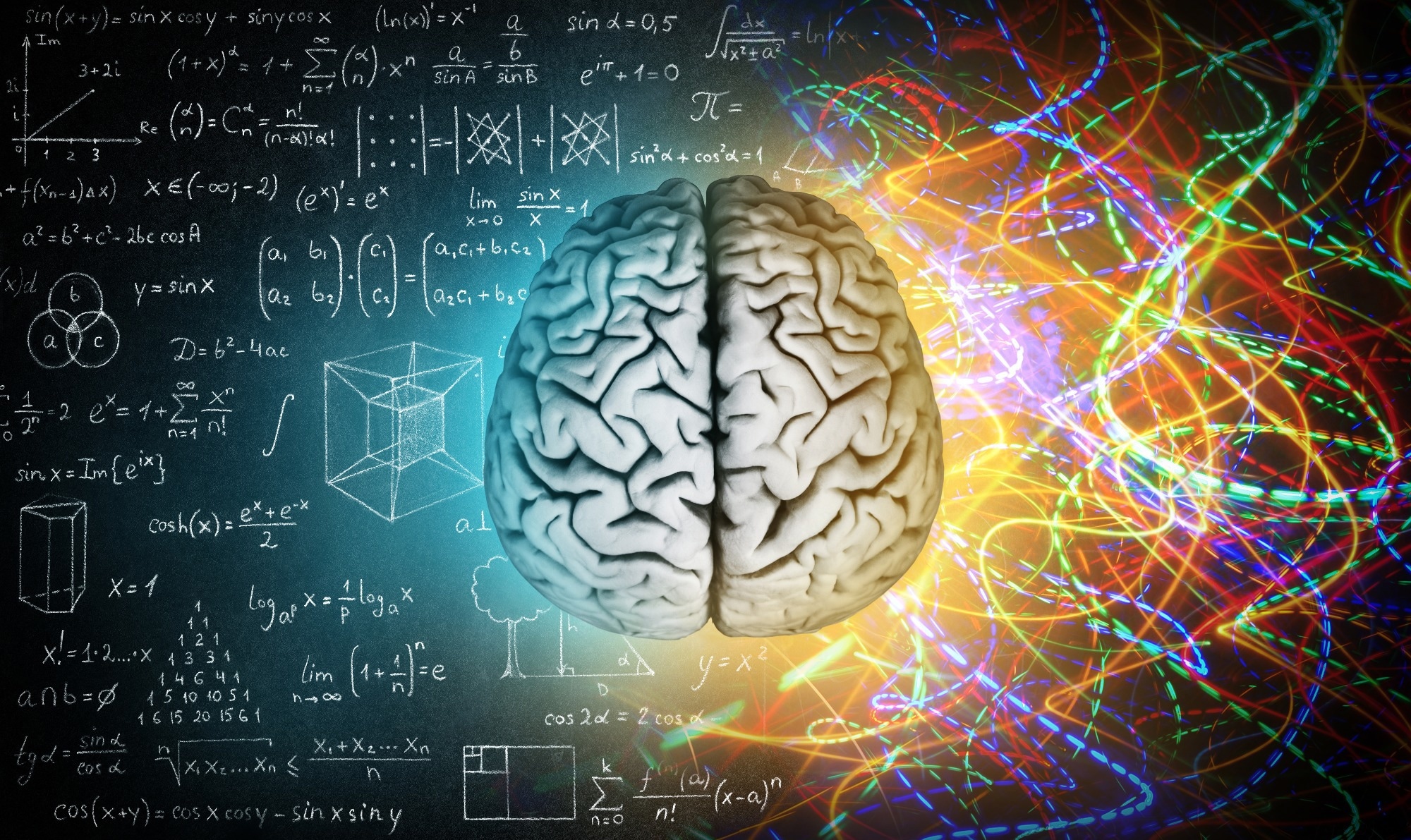 Image credit: Triff/Shutterstock.com
The human brain is among the most complex biological systems, composed of billions of neurons organized into interconnected networks that underpin cognition, behavior, and perception. A central goal of neuroscience has been to map this intricate “wiring diagram” to understand how structure gives rise to function and how its disruption contributes to disease.
This goal has evolved into the field of connectomics, a comprehensive “global” map of all neural connections within the nervous system that seeks to reconstruct neural connections across scales, from individual synapses between neurons to whole-brain networks.
DOWNLOAD the full PDF to uncover how connectomics is transforming our understanding of cognition, aging, and disease.
The Early Quest to Map the Brain
Early efforts to understand brain organization relied on anatomical observation. In the nineteenth century, the brain was widely viewed as a continuous “plumbing network”. This perspective changed when Santiago Ramón y Cajal demonstrated that the brain is composed of discrete cells called neurons, connected through specialized junctions called synapses. Cajal conceptualized the brain as a computational system composed of individual cellular units, whose intrinsic biophysical properties enable them to process and transmit information that gives rise to thought, emotion, and behavior.1
This early recognition that connections are essential for function remains the foundation of both modern connectomics and artificial neural networks. Today, scientists have built on this framework to learn how these remarkable cells relay information via electrochemical signaling rather than purely “zapping” electrical signals, enabling thought, movement, and communication.1
From Brain Regions to Neural Circuits
Twentieth-century neuroscience initially focused on mapping brain regions and associating them with specific functions. However, it became clear that brain activity arises from distributed networks rather than isolated areas. Functional imaging studies demonstrate that even simple cognitive processes recruit multiple interconnected regions, underscoring the importance of circuit-level organization.
The impact of this perspective is so significant that some neuroscientists now categorize the field's history as "Before Connectome" (BC) and "After Connectome" (AC). The first complete connectome was achieved in 1986 (published in full in 1992) for the nematode Caenorhabditis elegans, chosen for its simplicity with only 302 neurons. This Herculean effort took over a decade and involved serial-section electron microscopy and manual reconstruction from micrographs.2
Research@ NYC: Mapping the Brain
Video credit: GoogleResearch/Youtube.com
The Rise of Connectomics Technologies
Technological advances have been central to scaling connectomics. Electron microscopy (EM) provides the resolution required to identify synapses and reconstruct neural circuits at the nanometre scale. However, these methods generate extremely large datasets; even a cubic millimeter of human cortex can produce petabytes of data.4
To process this, researchers developed automated segmentation algorithms such as flood-filling networks (FFN). These AI systems function like pouring digital paint into a pipe, automatically tracing a neuron's volume across millions of image tiles. A key example is the reconstruction of a 1 mm³ volume of human temporal cortex, containing ~57,000 cells, hundreds of millimeters of vasculature, and ~150 million synapses. This dataset also revealed previously unrecognized neuronal cell types and rare, strong multisynaptic connections, demonstrating the power of large-scale connectomics.4
Whole-Brain Mapping Today
Modern whole-brain mapping integrates microscale and macroscale approaches. At the microscale, EM-based datasets provide detailed reconstructions of neural circuits. At larger scales, magnetic resonance imaging (MRI) enables non-invasive mapping of structural and functional connectivity across populations.
Large-scale imaging studies have produced normative “brain charts” that track structural variation across the human lifespan using datasets of over 120,000 MRI scans from more than 100,000 individuals. These frameworks allow individual brains to be interpreted within typical developmental and aging trajectories. Functional connectome analyses further reveal nonlinear patterns of change, with connectivity showing distinct developmental trajectories and system-specific maturation timelines rather than a single global peak.5-7
Advances are also extending connectomics beyond the brain. High-speed imaging methods now enable mapping of peripheral nerves at subcellular resolution across entire organisms within tens of hours, revealing interactions between neural and physiological systems. Similarly, PET-based approaches can generate whole-body connectomes that capture inter-organ metabolic communication using graph-based network models.8,9
 Image credit: Gorodenkoff/Shutterstock.com
Impact on Health and Disease Research
Connectomics is increasingly influencing the study of neurodegenerative and psychiatric disorders. By comparing individual connectivity patterns to normative datasets, researchers can identify deviations associated with disease. Large-scale analyses show that many disorders involve distributed, transdiagnostic patterns of structural and functional variation rather than isolated regional damage.5
Machine learning approaches enable researchers to integrate connectivity, imaging, and genetic data at scale. This supports the identification of biomarkers and improves understanding of disease mechanisms, helping to advance drug discovery and precision medicine approaches for complex neurological and psychiatric conditions.2
The Future of Brain Mapping
Despite rapid progress, major challenges remain. The scale of the human brain and the computational demands of high-resolution imaging continue to limit efforts to reconstruct a complete connectome. Artificial intelligence is expected to play a central role in addressing these challenges by enabling automated reconstruction and analysis of large datasets.2,4
Future directions also include expanding connectomics beyond the brain, reflecting the interconnected nature of neural and physiological systems. This aligns with earlier theoretical proposals that understanding behavior requires mapping interactions between the brain and the body as an integrated system.10
Connectomics aims not only to map the brain’s structure but to explain how its organization gives rise to cognition, behavior, and disease.
AI is not just about making computers smarter but making contributions to scientific questions like who we are and what we are.
Viren Jain, Google Research’s Connectomics Team lead researcher 3
As these efforts advance, they could transform neuroscience and medicine by enabling us to ask bigger questions, such as how consciousness and memories arise from neural connectivity.
References and Further Reading
- Pryor J. Who discovered neurons? MIT McGovern Institute. December 10, 2025. Accessed April 8, 2026. https://mcgovern.mit.edu/2025/12/10/who-discovered-neurons/
- Jain V. How AI could lead to a better understanding of the brain. Nature. 2023;623(7986):247-250. DOI:10.1038/d41586-023-03426-3, https://doi.org/10.1038/d41586-023-03426-3
- Google Research. Research@ NYC: Mapping the Brain. 2023. Accessed April 8, 2026. https://www.youtube.com/watch?v=aB_ZmAM3tv8
- Shapson-Coe A, Januszewski M, Berger DR, et al. A petavoxel fragment of human cerebral cortex reconstructed at nanoscale resolution. Science. 2024;384(6696):eadk4858. DOI:10.1126/science.adk4858, https://doi.org/10.1126/science.adk4858
- Bethlehem R a. I, Seidlitz J, White SR, et al. Brain charts for the human lifespan. Nature. 2022;604(7906):525-533. DOI:10.1038/s41586-022-04554-y, https://doi.org/10.1038/s41586-022-04554-y
- Sun L, Zhao T, Liang X, et al. Human lifespan changes in the brain’s functional connectome. Nat Neurosci. 2025;28(4):891-901. DOI:10.1038/s41593-025-01907-4, https://doi.org/10.1038/s41593-025-01907-4
- Taylor HP, Huynh KM, Thung KH, et al. Functional hierarchy of the human neocortex across the lifespan. Nature. Published online March 25, 2026:1-10. DOI:10.1038/s41586-026-10219-x, https://doi.org/10.1038/s41586-026-10219-x
- Shi MY, Yao Y, Wang M, et al. High-speed mapping of whole-mouse peripheral nerves at subcellular resolution. Cell. 2025;188(14):3897-3915.e20. DOI:10.1016/j.cell.2025.06.011, https://doi.org/10.1016/j.cell.2025.06.011
- Labarthe A, Varet S, Savale L, et al. Personalized mapping of body homeostasis using whole-body PET connectomics and routine FDG PET imaging. Commun Med. Published online March 27, 2026. DOI:10.1038/s43856-026-01549-y, https://doi.org/10.1038/s43856-026-01549-y
- Lo CC, Chiang AS. Toward Whole-Body Connectomics. J Neurosci. 2016;36(45):11375-11383. DOI:10.1523/JNEUROSCI.2930-16.2016, https://doi.org/10.1523/JNEUROSCI.2930-16.2016
Last Updated: Apr 9, 2026