Introduction
What Is Collagen Remodeling?
Why Collagen Architecture Matters
High-Resolution Imaging Techniques
Applications in Cancer and Fibrosis Research
Conclusion
References and Further Reading
Discover how high-resolution imaging techniques reveal structural changes in collagen associated with cancer and fibrosis, from microscopic fiber organization to molecular remodeling. Explore how these advances are improving our understanding of disease mechanisms and supporting the development of more precise diagnostic and therapeutic approaches.
 Image credit: nobeastsofierce/Shutterstock.com
Introduction
The extracellular matrix (ECM) is no longer viewed as a passive scaffold that simply supports tissues. Instead, it is increasingly recognized as a dynamic regulator of cell behavior, with collagen playing a central role in maintaining tissue architecture and influencing how cells communicate with their surroundings.1
In healthy tissues, collagen is continuously remodeled to preserve tissue structure and function. However, when this tightly regulated process becomes disrupted, the extracellular matrix undergoes progressive structural changes that alter tissue architecture and cell behavior. In cancer and fibrotic diseases, these alterations do more than reflect underlying pathology, they actively contribute to disease progression.2
Advances in high-resolution imaging are allowing researchers to visualize these structural changes with more detail, providing new insights into disease mechanisms and revealing potential biomarkers and therapeutic targets.3
This article explores how collagen remodeling contributes to cancer and fibrosis and how modern imaging technologies are reshaping our understanding of extracellular matrix biology.
What Is Collagen Remodeling?
Collagen is the most abundant structural protein in the extracellular matrix (ECM), providing tissues with strength, stability, and mechanical support. Throughout life, collagen is continuously renewed to support normal physiological processes such as growth, tissue repair, and wound healing while preserving the architecture and function of organs.2
This continual renewal, known as collagen remodeling, is a tightly regulated process that balances collagen production, organization, and degradation. Fibroblasts, the principal collagen-producing cells, synthesize procollagen, the soluble precursor of mature collagen. Once secreted into the extracellular space, procollagen molecules undergo enzymatic processing before assembling into collagen fibrils and larger fibers, whose orientation, density, and cross-linking determine the mechanical properties of tissues. Different collagen types contribute to distinct extracellular matrix compartments: fibrillar collagens, including types I and III, are prominent in the interstitial matrix, whereas type IV collagen forms networks within basement membranes.1,2,5
At the same time, enzymes known as matrix metalloproteinases (MMPs) degrade aged or damaged collagen so it can be replaced. Together, these coordinated processes maintain collagen homeostasis, ensuring that the extracellular matrix remains both structurally stable and adaptable to changing physiological demands. Collagen turnover also involves other proteolytic pathways, and the balance between matrix synthesis and degradation varies according to tissue type, physiological state, and injury response.1,2,6
Chronic injury, persistent inflammation, or disease can disrupt this balance by keeping fibroblasts activated and altering the normal turnover of collagen. As collagen production begins to outpace its degradation, newly synthesized fibers accumulate within the extracellular matrix. Over time, these fibers may become increasingly cross-linked and reorganized into denser, more aligned networks, fundamentally altering the architecture of the ECM. As a result, tissues become progressively stiffer, and the physical environment surrounding cells begins to change. However, pathological remodeling is not restricted to increased collagen production: impaired matrix degradation, excessive proteolysis, and abnormal collagen organization can also compromise tissue structure and function.1,2
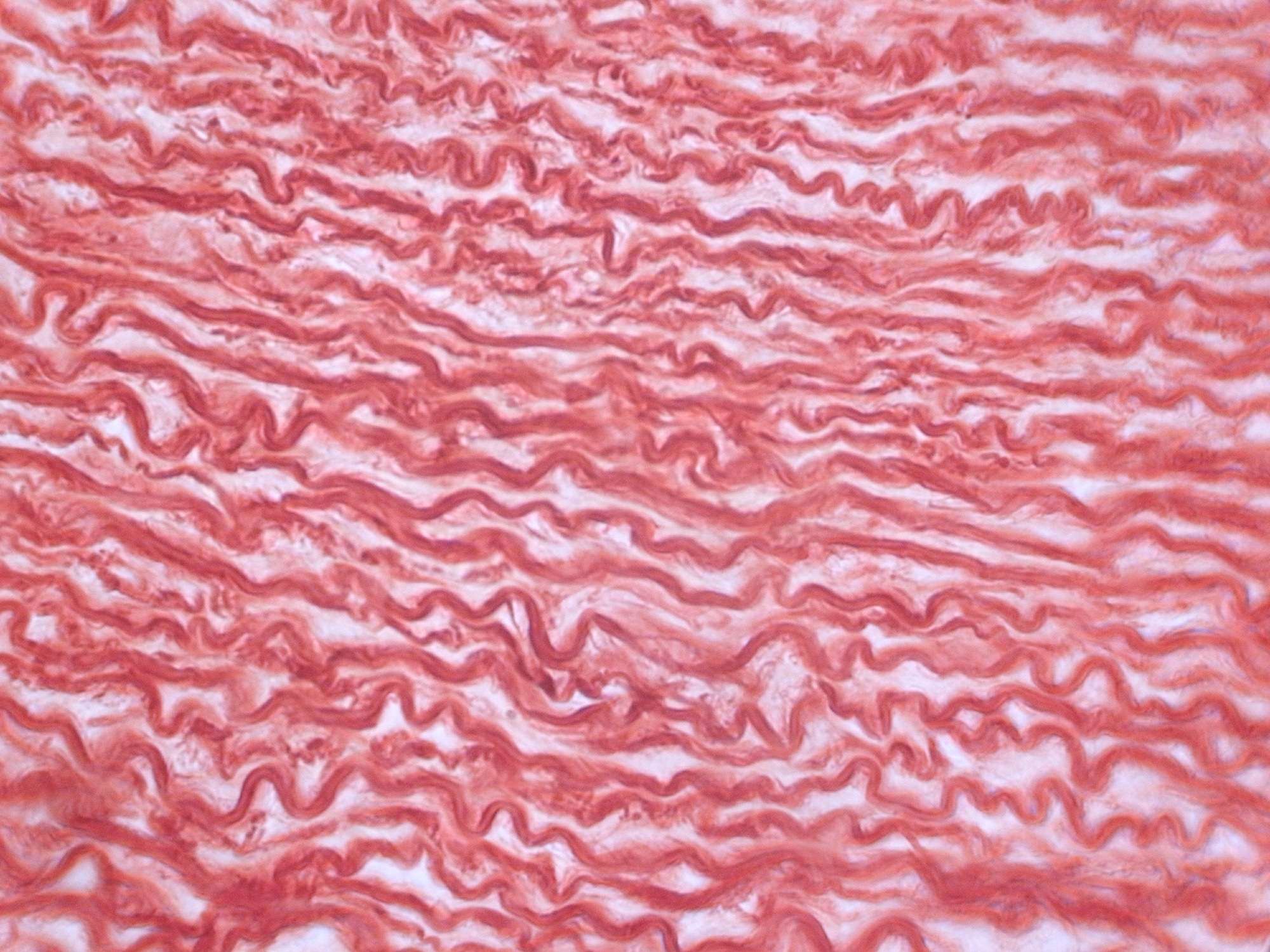 Microscopic view of densely arranged connective tissue fibers, illustrating the layered and undulating architecture of the extracellular matrix. Changes in collagen fiber organization and density are important features of tissue remodeling in cancer and fibrosis. Image credit: Anna Jurkovska/Shutterstock.com
Why Collagen Architecture Matters
The structural changes produced by collagen remodeling have significant biological consequences. As collagen fibers become denser, increasingly aligned, and more extensively cross-linked, the extracellular matrix can become stiffer, and its biochemical and mechanical properties change. Cells detect these alterations through mechanotransduction, the process by which mechanical forces are converted into biochemical signals, allowing collagen architecture to actively influence cell behavior. Integrin-mediated interactions between cells and the extracellular matrix help transmit these mechanical signals, while enzymes such as lysyl oxidases contribute to collagen cross-linking and changes in matrix stiffness.2,4,6
In cancer, these architectural changes create a microenvironment that can actively support disease progression. Many fibroblasts adopt an activated phenotype known as cancer-associated fibroblasts (CAFs), which continually remodel the extracellular matrix through collagen deposition and reorganization. Collagen remodeling is also influenced by cancer cells, immune cells, and other stromal populations, making the tumor microenvironment a dynamically regulated system rather than a passive accumulation of fibrous tissue.1,4,5
The resulting aligned collagen fibers can create physical pathways that facilitate cancer-cell migration, while matrix stiffening activates mechanotransduction pathways that promote cell survival, proliferation, angiogenesis, and invasion. Collagen remodeling also alters immune-cell infiltration and function, potentially contributing to an immunosuppressive microenvironment that supports tumor progression. Together, these interactions establish a positive feedback loop in which cancer cells and the remodeled extracellular matrix reinforce one another. However, collagen's effects are context-dependent: particular collagen types and structural arrangements may also restrict tumor expansion, and extensive matrix deposition can hinder drug penetration and immune-cell access.1,4,5
In fibrotic diseases, collagen remodeling follows a different but equally damaging trajectory. Following chronic injury, fibroblasts and activated myofibroblasts can remain persistently activated and continue producing collagen long after normal tissue repair should have ended. Progressive collagen deposition and cross-linking replace healthy tissue with dense scar-like matrix, increasing tissue stiffness and disrupting normal organ architecture. Increased matrix stiffness can, in turn, sustain myofibroblast activation, reinforcing a cycle of abnormal matrix production and mechanical signaling. This process underlies diseases such as liver fibrosis, idiopathic pulmonary fibrosis, and oral submucous fibrosis, all of which are characterized by excessive collagen accumulation and impaired tissue function. In some cases, prolonged fibrosis also creates a tissue environment that increases the risk of cancer development. Although fibrosis and cancer share mechanisms involving inflammation, fibroblast activation, and extracellular matrix remodeling, their progression and clinical consequences differ across organs.3,6
Because these architectural changes closely reflect aspects of disease activity, collagen organization has emerged as an important candidate diagnostic and prognostic biomarker. Quantifying features such as collagen fiber orientation, density, and organization is helping researchers better characterize disease progression, evaluate therapeutic responses, and identify new targets for precision medicine. Nevertheless, collagen architecture alone cannot reliably distinguish active matrix formation from established fibrosis, because similar structural appearances may arise from different rates of collagen synthesis, degradation, and cross-linking.2,6
High-Resolution Imaging Techniques
The structural changes associated with collagen remodeling are difficult to fully capture using conventional histology. Although routine staining can reveal collagen deposition, it provides limited information about collagen fiber organization, orientation, and three-dimensional architecture. High-resolution imaging techniques can help overcome these limitations by enabling researchers to visualize and quantify collagen remodeling in far greater detail. However, spatial resolution, imaging depth, molecular specificity, sample preparation, and the ability to examine living tissues vary substantially between techniques.3,9
Among these methods, second harmonic generation (SHG) microscopy has become one of the most widely used techniques for studying fibrillar collagen. SHG exploits collagen's intrinsic optical properties to generate high-resolution, label-free images of collagen fibers, making it particularly valuable for quantifying fiber orientation, density, and organization in intact tissues. The technique relies on a nonlinear optical interaction with ordered, non-centrosymmetric molecular structures and is particularly sensitive to fibrillar collagen organization. However, because SHG selectively detects highly ordered fibrillar collagen, it provides limited information about non-fibrillar matrix components and surrounding cells. Importantly, SHG signal intensity should not be interpreted as a direct measurement of collagen concentration, because it also depends on fibril organization, orientation, optical polarization, and tissue scattering. Polarization-resolved and directional SHG approaches can provide additional information about collagen organization.7
Video 2 SHG 3D imaging of collagen fibers
Video credit: @TheWarrenAlpertMedicalSchool/Youtube.com
Multiphoton microscopy provides a broader imaging framework that can incorporate SHG alongside two-photon excited fluorescence and other nonlinear optical signals. These approaches commonly employ near-infrared excitation, enabling optical sectioning and imaging at greater depths than many conventional fluorescence methods, depending on tissue properties and instrumentation. As a result, researchers can visualize collagen remodeling within intact tissues while preserving much of the native tissue environment, although the technique requires specialized instrumentation. Combining SHG with endogenous two-photon autofluorescence can reveal fibrillar collagen alongside cellular and other tissue features, while fluorescence lifetime measurements can provide complementary information about tissue metabolism. Imaging depth and photodamage remain dependent on excitation conditions and specimen characteristics.7,8,9
To build a more complete picture of collagen remodeling, researchers also use confocal, super-resolution, and electron microscopy, each providing complementary information about collagen architecture and its interactions with surrounding cells. Confocal microscopy enables optical sectioning and three-dimensional reconstruction, allowing collagen to be visualized alongside fluorescently labeled cells and proteins. Its imaging depth, however, is more limited than multiphoton microscopy. Unlike label-free SHG imaging of fibrillar collagen, fluorescence-based confocal imaging generally requires suitable labels or endogenous fluorescent contrast to distinguish specific extracellular matrix components.9
Super-resolution microscopy uses approaches such as structured illumination microscopy, stimulated emission depletion microscopy, and single-molecule localization microscopy to achieve spatial resolution beyond conventional diffraction-limited fluorescence imaging. These techniques can reveal selected nanoscale features of collagen organization and matrix–cell interactions when suitable labeling and imaging conditions are available. However, its broader application remains constrained by specialized instrumentation, technical complexity, and challenges associated with imaging living tissues. Reported resolution must be interpreted carefully, because localization precision, reconstructed image appearance, and the ability to distinguish nearby biological structures are not equivalent. Labeling density, image processing, specimen movement, and acquisition conditions can all influence the effective resolution and reliability of the resulting images.9,10
At even higher resolution, electron microscopy provides ultrastructural visualization of individual collagen fibrils and fiber organization. Transmission electron microscopy (TEM), in particular, enables examination of fibril cross-sections, allowing quantitative assessment of properties such as fibril cross-sectional area and aspect ratio. Semi-automated image segmentation and statistical analysis can reduce subjectivity and improve the consistency of these measurements. However, because conventional electron microscopy is typically performed on fixed specimens, it cannot directly visualize dynamic biological processes in living tissues. Its quantitative interpretation also depends on specimen preparation, section orientation, image quality, and the representativeness of sampled tissue regions.11,12
Because no single imaging modality can fully characterize collagen remodeling, researchers are increasingly combining complementary approaches through correlative and multimodal imaging. By integrating structural, molecular, and functional information from the same specimen, these approaches provide a more comprehensive view of extracellular matrix organization and remodeling. For example, SHG can characterize fibrillar collagen architecture, fluorescence-based methods can identify cellular or molecular features, and electron microscopy can examine fibril ultrastructure. Combining these measurements requires careful image registration and attention to differences in specimen preparation and spatial scale.9,11,13
Correlated multimodal imaging specifically aims to relate measurements from corresponding specimens and regions of interest. Accurate registration, specimen relocation, compatible preparation protocols, and management of large multidimensional datasets are therefore central methodological challenges.13
Advances in computational image analysis and artificial intelligence are further expanding the quantitative information that can be extracted from these datasets, supporting the development of imaging biomarkers with potential applications in disease diagnosis, prognostic assessment, and treatment monitoring. Automated measurements nevertheless require validation against appropriate reference methods and assessment of reproducibility before they can be used reliably across laboratories or clinical settings.3,11,14
Applications in Cancer and Fibrosis Research
High-resolution imaging is providing new insights into how collagen remodeling influences disease progression. In cancer, researchers are using these techniques to characterize the tumor microenvironment by quantifying collagen fiber orientation, density, and organization, features that are increasingly associated with tumor aggressiveness, metastatic potential, and, in some settings, response to immunotherapy.15
In breast cancer, second harmonic generation (SHG) microscopy has shown that aligned collagen fibers form distinct patterns known as Tumor-Associated Collagen Signatures (TACS). These patterns are associated with increased tumor invasion and poorer clinical outcomes, demonstrating how collagen architecture can provide prognostic information beyond conventional histopathology. Collagen density and alignment have also been associated with clinical outcomes, although findings vary across studies and tumor subtypes.16
In cancer immunotherapy, dense collagen networks can restrict immune-cell infiltration and contribute to treatment resistance, highlighting collagen remodeling as a potential therapeutic target.15
In fibrotic diseases, quantitative imaging is helping researchers assess collagen deposition and tissue remodeling to monitor disease progression and evaluate treatment response. In idiopathic pulmonary fibrosis (IPF), for example, collagen hybridizing peptides (CHPs) can detect denatured collagen associated with active matrix remodeling. Experimental fluorescence and positron emission tomography (PET) imaging using CHPs has shown promise for assessing pulmonary fibrosis, although clinical validation is still required.17
These advances illustrate how quantitative analysis of collagen architecture is improving researchers' understanding of disease biology while supporting the development of more effective diagnostic and therapeutic strategies. However, standardized imaging methods and clinical validation remain essential before these approaches can be routinely applied in patient care.14
Conclusion
High-resolution imaging has transformed the study of collagen remodeling by enabling researchers to move beyond qualitative observation of the extracellular matrix toward quantitative analysis of tissue architecture. By revealing how collagen fibers are organized, aligned, and remodeled throughout disease progression, these technologies are providing new insights into the biological processes that drive cancer and fibrosis. Importantly, different imaging modalities reveal different aspects of collagen biology, and structural measurements should not automatically be interpreted as direct indicators of collagen concentration, mechanical stiffness, or active matrix turnover.2,7,9
As imaging continues to evolve alongside advances in molecular biology and computational analysis, it is expected to provide a more integrated understanding of the extracellular matrix. This deeper understanding may ultimately help researchers identify more robust biomarkers, evaluate emerging therapies, and translate advances in collagen biology into improved patient care. Achieving this goal will require standardized acquisition and analysis methods, integration of complementary imaging and molecular measurements, and rigorous validation of candidate biomarkers in clinically relevant settings.3,9
In particular, combining quantitative collagen architecture with molecular indicators of collagen degradation and turnover may help distinguish established matrix accumulation from ongoing remodeling, while carefully validated imaging
References and Further Reading
- Song, K. et al. Collagen Remodeling along Cancer Progression Providing a Novel Opportunity for Cancer Diagnosis and Treatment. Int. J. Mol. Sci. 23, 10509 (2022). DOI:10.3390/ijms231810509, https://www.mdpi.com/1422-0067/23/18/10509
- Mayorca-Guiliani, A. E. et al. ECM formation and degradation during fibrosis, repair, and regeneration. Npj Metab. Health Dis. 3, 25 (2025).
- Baniasadi, A. et al. Imaging at the nexus: how state of the art imaging techniques can enhance our understanding of cancer and fibrosis. J. Transl. Med. 22, 567 (2024).
- Ashworth, J. C. & Cox, T. R. The importance of 3D fibre architecture in cancer and implications for biomaterial model design. Nat. Rev. Cancer 24, 461–479 (2024).
- Zhou, Y., Jiang, Z., Cao, L. & Yang, J. The role of various collagen types in tumor biology: a review. Front. Oncol. 15, 1549797 (2025). DOI:10.3389/fonc.2025.1549797, https://www.frontiersin.org/journals/oncology/articles/10.3389/fonc.2025.1549797/full
- Rieder, F. et al. Fibrosis: cross-organ biology and pathways to development of innovative drugs. Nat. Rev. Drug Discov. 24, 543–569 (2025).
- Aghigh, A. et al. Second harmonic generation microscopy: a powerful tool for bio-imaging. Biophys. Rev. 15, 43–70 (2023).
- Chen, K., Han, Y., Wang, Z. & Cui, Y. Submicron resolution techniques: Multiphoton microscopy in skin disease. Exp. Dermatol. 32, 1613–1623 (2023).
- Poole, J. J. A. & Mostaço-Guidolin, L. B. Optical Microscopy and the Extracellular Matrix Structure: A Review. Cells 10, 1760 (2021). DOI:10.3390/cells10071760, https://www.mdpi.com/2073-4409/10/7/1760
- Prakash, K. et al. Resolution in super-resolution microscopy, definition, trade-offs and perspectives. Nat. Rev. Mol. Cell Biol. 25, 677–682 (2024).
- Rego, B. V., Weiss, D. & Humphrey, J. D. A Fast, Robust Method for Quantitative Assessment of Collagen Fibril Architecture from Transmission Electron Micrographs. Microsc. Microanal. 29, 2099–2107 (2023).
- Hart, R. C., Carroll, D. T., Vinogradova, M. & Krystofiak, E. S. A Guide to Fixation and Staining of Biological Samples for Electron Microscopy. Microsc. Today 32, 37–40 (2024).
- Rudraiah, P. S. et al. Correlated multimodal imaging in life sciences: lessons learnt. Front. Biomater. Sci. 3, 1338115 (2024). DOI:10.3389/fbiom.2024.1338115, https://www.frontiersin.org/journals/biomaterials-science/articles/10.3389/fbiom.2024.1338115/full
- Eertink, J. J. et al. The development process of ‘fit-for-purpose’ imaging biomarkers to characterize the tumor microenvironment. Front. Med. 11, 1347267 (2024). DOI:10.3389/fmed.2024.1347267, https://www.frontiersin.org/journals/medicine/articles/10.3389/fmed.2024.1347267/full
- Borst, R., Meyaard, L. & Pascoal Ramos, M. I. Understanding the matrix: collagen modifications in tumors and their implications for immunotherapy. J. Transl. Med. 22, 382 (2024).
- Heydari, S. et al. The association between tumor-stromal collagen features and the clinical outcomes of patients with breast cancer: a systematic review. Breast Cancer Res. 27, 69 (2025).
- Zhao, J. et al. Delineating, Imaging, and Assessing Pulmonary Fibrosis Remodeling via Collagen Hybridization. ACS Nano 18, 27997–28011 (2024).
Last Updated: Oct 8, 2026