Overview
EpiVaginal offers an in vitro research tool for drug development, feminine product safety testing, and the study of HIV-1 and other sexually transmitted diseases. Human-like 3D tissue structure and cellular physiology make it ideal for toxicology, drug delivery, and formulation efficacy testing.
Its cellular physiology and tissue structure are nearly similar to in vivo vaginal epithelial tissue, thereby making it perfect for toxicology and efficacy testing, as well as a tool that has been utilized for biological research.
Technology
EpiVaginal tissues have been cultured from normal, human-derived vaginal epithelial, and dendritic cells. Its highly-differentiated structure matches in vivo tissue and is perfect for toxicity studies of vaginal care, feminine hygiene, and microbicide products. Four kinds of EpiVaginal are provided.
1) VEC-100 (Figure 1): An epithelial tissue consisting of epithelial VEC cells
2) VLC-100 (Figure 1): An epithelial tissue containing epithelial VEC and immuno-competent dendritic cells,
3) VEC-100-FT (Figure 2): A full-thickness version of VEC-100 including VEC epithelial cells and a fibroblast-containing lamina propria, and
4) VLC-100-FT (Figure 2): An immuno-competent version of the VEC-100-FT which consists of dendritic cells.
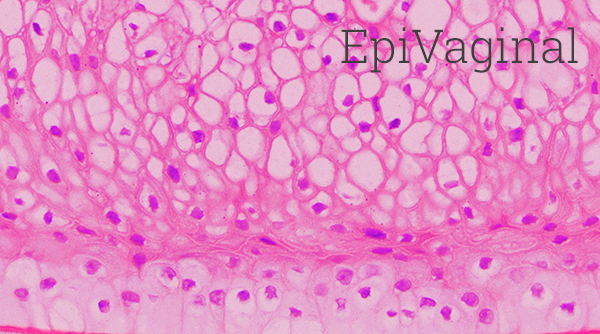
Figure 1. H&E stained histological (formalin-fixed) cross-sections of A) VEC-100 in vitro reconstructed epithelial tissue models containing normal human VEC cells, and B) vaginal explant tissue. Both in vitro and in vivo tissues show nucleated basal and suprabasal cell layers followed by layers in which nuclei are lost and cells are filled with glycogen. Image Credit: MatTek
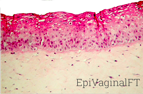
Figure 2. H&E stained histological (formalin-fixed) cross-sections of full-thickness EpiVaginal tissues. The VEC-100-FT tissues consist of VEC epithelial cells cultured atop a lamina propria (LP)-like collagen matrix that contains fibroblasts. The VLC-100-FT consists of VEC epithelial and dendritic cells in the epithelial layers and contains both fibroblasts and dendritic cells in the LP. Image Credit: MatTek
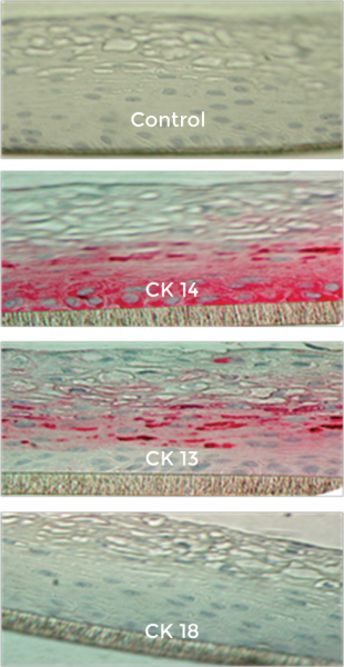
Figure 3. Formalin-fixed cross-sections of the VEC-100 tissue immuno-stained for cytokeratins, CK13, CK14, and CK18. Basal cells are stained by CK14 and supra-basal cells by CK13; no cells are stained by CK18. Image Credit: MatTek
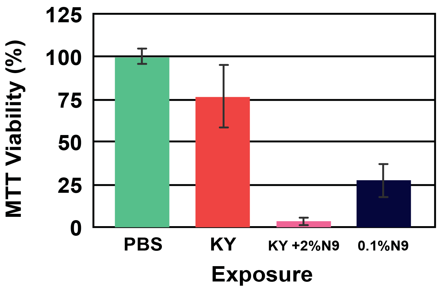
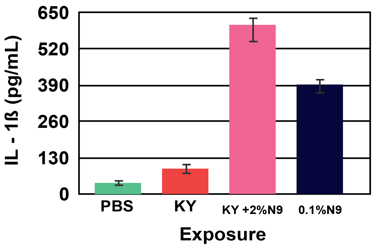
Figure 4. Viability (A) and Cytokine Release (B) of EpiVaginal full-thickness (VEC-100-FT) tissue model following 18 hour exposure to formulations containing the common spermicide, nonoxynol-9 (N9): a) PBS control, b) KY jelly (KY), c) KY with 2% N9 (2% N9), and d) 0.1% N9 in PBS (0.1% N9). Note: As the tissue viability decreases, the IL-1β release increases. Image Credit: MatTek
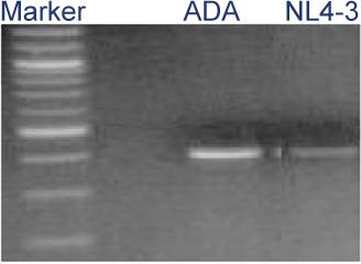
Figure 5. HIV-1 infection of VLC-100 tissue following topical exposure (24hr) to 60,000 CPM of HIV-1 ADA or NL4-3 virus. After exposure, tissues were washed 3X and then cultured for an additional 24 hr. Cellular DNA was then extracted and HIV-1 transcripts were detected using HIV-1 gag specific primer pairs. Image Credit: MatTek

Figure 6. RT PCR showing Toll-like Receptor (TLR) expression for: A) organotypic vaginal-ectocervical (VEC) tissue model and B) Human cervical tissue explant. Image Credit: MatTek
Human relevance
The variability and late-stage failures of in vivo testing can be prevented. It is possible for users to assess the irritancy of surfactant-based vaginal products in the developmental stage, with the help of an EpiVaginal and a simple in vitro assay.
Applications
- STD Infection research
- Microbicide testing
- Inflammation
- Feminine hygiene
- Drug delivery
Irritation testing
Utilize the EpiVaginal tissue to quantify the tissue viability and cytokine release after exposure to feminine hygiene and other products.
EpiVaginal irritation protocol
EpiVaginal offers an in vitro research tool both available for feminine product safety testing and the study of HIV-1 as well as other sexually transmitted diseases.
The cellular physiology and tissue structure are closely parallel to in vivo vaginal epithelial tissue, thereby making it perfect for efficacy and toxicology testing, as well as a tool for biological research.