Overview
An in vitro model of melanoma provides cancer researchers with a deep insight into disease progression. It is also an effective platform for new therapies to be tested.
Technology
The Melanoma model comprises human malignant melanoma cells (A375), normal, human-derived epidermal keratinocytes (NHEK), and normal, human-derived dermal fibroblasts (NHDF) which have been cultured to develop a multilayered, highly distinguished epidermis with melanoma cells at several stages of CM malignancy.
At various stages of the culture, the tissue displays radial growth phase (RGP), vertical growth phase (VGP), or metastatic melanoma phenotype. The cells have been cultured on cell culture inserts with the help of a serum-free medium, and obtain levels of differentiation on the cutting edge of in vitro skin technology
In a structural manner, the Melanoma model nearly parallels the advancement of melanoma in vivo, thus offering a useful tool to study, comprehend, and develop preventative and therapeutic treatments for one of the highly serious cutaneous malignancies.
The Melanoma model displays in vivo-like morphological and growth characteristics which are even and highly reproducible. The epidermis of this full-thickness skin model comprises organized basal, granular, spinous, and cornified epidermal layers analogous to those found in vivo. The dermal compartment is made of a collagen matrix consisting of viable normal human dermal fibroblasts (NHDF).
The protocols for utilizing the Melanoma tissues are clear and direct. MatTek’s Melanoma tissues have been used with several targets and anti-melanoma drugs.
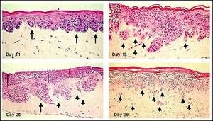
Figure 1. Human Metastatic Melanoma Cells (A375) in Full Thickness Melanoma Skin Model. A375 cells develop RGP melanoma nodes at dermal/epidermal junction (Day 11). With extended culture time, melanoma nodes adopt a VGP morphology (Day 18) and subsequently isolated clusters of cells invade the dermis (metastatic invasion) (Day 29). Long arrows indicate melanoma cell clusters at the epidermal-dermal junction. Short arrows show separated melanoma cell clusters infiltrating the dermis. Image Credit: MatTek
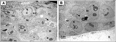
Figure 2. Ultrastructural analysis of Melanoma FT Skin Model. Transmission electron micrograph (TEM) of the full thickness skin melanoma model (MLNM-FT-A375). A. Area of interaction of melanoma cells (M) and keratinocytes (KC). B. Area of interaction of melanoma cells and the underlying dermal substrate (D). N, nucleus, n, nucleoli. Arrow indicates apoptotic melanoma cells. Image Credit: MatTek
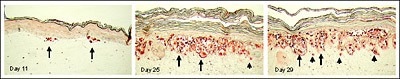
Figure 3. Full thickness skin melanoma model (MLNM-FT) containing metastatic SK-Mel-28 cells. S-100 antibody staining. Initially small nests of melanoma cells form at the dermal/epidermal junction (Day 11). Later VGP tumors develop (Day 25), and subsequently metastasis into the dermis is observed (Day 29). Long arrows indicate some of the melanoma cell clusters at the epidermal-dermal junction. Short arrows show individual melanoma cells infiltrating the dermis. Image Credit: MatTek
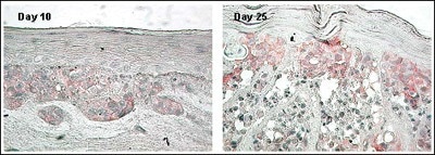
Figure 4. Expression of N-Cadherin Adhesion Molecule in MLNM-FT-A375 Full Thickness Melanoma Skin Model. N-Cadherin antibody staining, 40×. Note increased level of N-Cadherin expression with time in culture. Image Credit: MatTek
Human relevance
MatTek’s melanoma model matches the morphology and evolution of melanoma in vivo, making it an invaluable tool for oncological research and the evaluation of therapeutic candidates’ effectiveness.
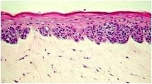
Image Credit: MatTek
Applications
Tumor invasion and anti-melanoma drug screening
MatTek’s Melanoma tissue displays a vertical growth phase, radial growth phase, and metastatic melanoma phenotypes, paralleling the advancement of melanoma in vivo.
This model offers a beneficial tool for oncologists and cancer scientists to study, comprehend, and come up with preventative and therapeutic treatments for one of the most serious cutaneous malignancies.