Triple-negative breast cancer carries the poorest prognosis among breast cancer subtypes, partly because of its high metastasis rate. Emerging research suggests that tumor-infiltrating nerves contribute to the aggressiveness of these cancers, but how tumor tissue recruits nerves remains unclear.
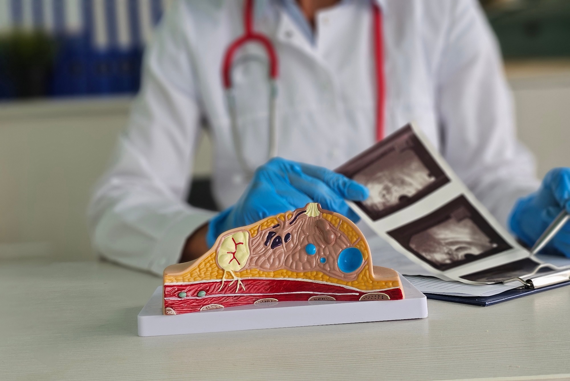 Study: Macrophage-secreted brain-derived neurotrophic factor promotes tumor growth in triple-negative breast cancer by inducing axonogenesis. Image Credit: NMK-Studio/Shutterstock.com
In a recent study published in Cell Death & Differentiation, researchers found that tumor-associated macrophages supply the molecular signal needed to recruit nerves into breast tumors.
Background
Triple-negative breast cancer lacks the hormone receptors that guide many other treatment strategies, leaving patients with fewer treatment options and a higher risk of relapse. The tumor microenvironment (TME), consisting of blood vessels, stromal cells, nerves, and immune cells, shapes how the tumor progresses or responds to treatment. Growing research suggests that nerves are active participants in the TME rather than passive bystanders.
Sensory nerves in particular are abundant in triple-negative tumors, and evidence from other cancers suggests they can dampen the immune system's ability to fight malignant cells. Tumors are thought to recruit these nerves through axonogenesis, where released growth factors signal nearby neurons to extend new axons into the tumor tissue. However, the molecular mechanisms of axonogenesis in the TME are not well understood.
Want to save this article for later? Click here.
The Study
Researchers used a mouse model of triple-negative breast cancer to determine whether immune cells supply the brain-derived neurotrophic factor (BDNF) needed for tumor innervation. The study used mice genetically engineered to lack BDNF specifically in immune cells, along with genetically matched littermates as controls.
All mice received transplants of breast cancer cells into the mammary fat pad. The researchers tracked tumor growth over several weeks and examined harvested tumors for grade and volume. Tumor sections were then stained with antibodies against pan-neuronal, sympathetic, and sensory nerve markers; fluorescence microscopy was used to visualize how densely nerves infiltrated tumors.
To test whether sensory nerves were specifically required for tumor growth, the team chemically eliminated nerves carrying a heat- and capsaicin-sensitive receptor before transplanting two different triple-negative tumor cell lines, and compared these outcomes to genetic elimination of the same nerve population in a separate cohort.
To identify which immune cells produce the neurotrophic factor, the researchers collected tumors at multiple time points after transplant and measured gene expression and protein levels. The researchers also cultured bone marrow-derived macrophages in media conditioned by tumor cell lines of differing aggressiveness to observe how tumor signals shaped macrophage behavior.
To determine whether macrophage-derived signaling alone was sufficient to restore tumor growth, macrophages from normal and deficient mice were transplanted into hosts of each genotype, and tumor establishment and innervation were assessed.
Lastly, the team analyzed human breast cancer datasets from a publicly available genomic repository to examine associations between macrophage presence, gene activity, and patient survival.
Key Findings
Macrophages served as the primary source of the signal that recruited nerves into triple-negative breast tumors.
Mice lacking BDNF in their immune cells largely failed to develop tumors after transplantation, and the few tumors that did form were smaller and less advanced than those in normal littermates. These tumors also showed substantially reduced nerve infiltration, with significantly fewer sensory nerves than sympathetic ones.
Depleting sensory nerves directly, either chemically or through genetic modification, slowed tumor growth across multiple mouse models, emphasizing that nerve infiltration actively supports tumor expansion rather than simply accompanying it.
Macrophages were also responsible for most of the BDNF produced in the TME, and the expression of BDNF was found to peak early after tumor formation before declining. The study also found that macrophages exposed to signals from a more aggressive tumor line were more likely to adopt a tumor-supportive profile, whereas exposure to a less aggressive line did not trigger the same shift.
The researchers observed that transferring healthy macrophages capable of producing BDNF into deficient mice restored both tumor growth and nerve infiltration, confirming that this immune signal alone can drive the process. Conversely, blocking the nerve receptor that responds to BDNF also reduced tumor size and nerve density in two separate models.
In datasets drawn from human breast cancer patients, higher macrophage presence combined with higher activity of this signaling pathway corresponded with worse survival outcomes, suggesting that the mechanism observed in mice may carry clinical relevance. However, the authors noted that other cell types, including tumor cells themselves and surrounding connective tissue cells, can also produce this neurotrophic factor, which may explain why some nerve infiltration persisted even when the immune-derived source was absent. This suggests multiple, potentially overlapping pathways contribute to tumor innervation.
Conclusions
The key findings from this study indicated that macrophages are a critical source of the signal that recruits nerves into triple-negative breast tumors and suggested that this immune-driven process actively supports tumor growth.
Observations from both mouse models and human data link macrophage activity, nerve infiltration, and disease severity in triple-negative breast cancers, and indicate that the BDNF signaling pathway could be a potential therapeutic target for a cancer subtype that currently offers patients few treatment options.
Journal reference:
Abbadi, J., Velayutham, R., Annan, A. C., Nikpoor, A. R., Ahmadi, M., Rocha, B. G. S., Farriester, J. W., Reel, J. M., Holland, E. C., Szulzewsky, F., Birbrair, A., Fung, K.-M., Talbot, S., & Cox, M. A. (2026). Macrophage-secreted brain-derived neurotrophic factor promotes tumor growth in triple-negative breast cancer by inducing axonogenesis. Cell Death & Differentiation. DOI:10.1038/s41418-026-01796-5
https://www.nature.com/articles/s41418-026-01796-5
 Single-Cell Metabolic Profiling Identifies Key Bioenergetic Targets in Human Tumor Tissue