The desire to develop smaller and smaller devices and enable smaller devices to have enhanced capability has meant that we need to see smaller and smaller components. Optical microscopy has improved with greater magnification and reduced aberrations, but this can only achieve a maximum magnification of around one thousand times. This level of magnification is not enough for the demands of modern technology.
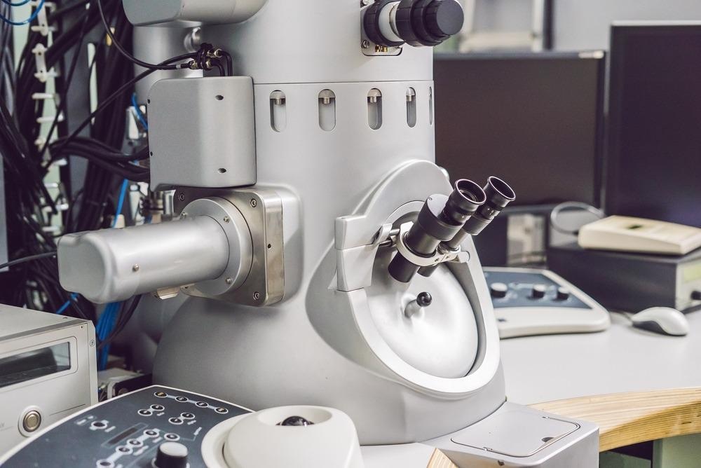
Image Credit: Elizaveta Galitckaia/Shutterstock.com
Electron microscopy can provide much greater magnification than optical microscopy, and this enhanced performance is because of the shorter wavelength of an electron than a photon. A scanning electron microscope (SEM) will allow up to 2 million times magnification and a transmission electron microscope (TEM) up to 50 million times with resolution down to half an angstrom (0.5 x 10⁻¹⁰metres) which is atomic scale. This massive magnification has vast implications for discovering and confirming facts about materials. There are limitations to this powerful technique as viewing samples at such high magnification may require special sample preparation.
It is now possible to see the structures of life forms as small as viruses with a Transmission electron microscope. This enhanced capability has massive implications for advancing life sciences and materials and environmental sciences.
What is a Transmission Electron Microscope
A scanning electron microscope can be as small as desktop size, but a Transmission microscope is a more substantial piece of equipment. A Transmission Electron Microscope also needs a controlled environment to function correctly, and a Transmission Electron Microscope requires a vibration-free environment with no electric or magnetic fields.
A TEM will also need a significant electric supply and will likely need a cooling source to prevent the machine from overheating. These requirements mean that a large budget is required to install this type of equipment, typically hundreds of thousands of dollars. Manufacturers like Zeiss, Jeol, Philips, Thermo Fisher, and Hitachi can supply Transmission Electron Microscopes and other related equipment.
A transmission Electron Microscope operates in a similar way to an optical microscope. A typical TEM construction consists of:-
- Electron beam
- Electromagnetic lenses
- Condensers
- Sample stage
- Phosphor fluorescent screen
- Computer screen
- All components are in a vacuum chamber.
The electron source is usually a tungsten or Lanthanum hexaboride filament with a high voltage, typically 60 – 300 kV applied to it. The filament releases electrons which are focussed into a beam and accelerated towards the sample stage, and some will pass through the sample, while some will not. Electrons transmitted through the sample produce a high-resolution two-dimensional black and white image on a fluorescent screen which is recorded on a computer.
Because the electrons need to pass through a sample is has to be “electron transparent,” which means it has to be very thin, typically less than 100 nanometers thick. This requirement means that sample preparation can be complex and requires specially trained staff, and samples may require additional treatment in addition to being sliced very thin.
It is possible to combine a Scanning Electron Microscope with a Transmission Electron Microscope in a Scanning Transmission Electron Microscope (STEM) to provide an even greater amount of detail about the sample. High-Resolution Transmission Electron Microscopy (HRTEM)is used to study materials down to atomic size.
Sample Preparation
Sample preparation for Transmission Electron Microscopy may require several specialized techniques. Biological samples usually require one of two methods to make them suitable for slicing very thin
Chemical fixation – chemicals, such as glutaraldehyde are used to crosslink proteins to stabilize them.
Cryofixation – where a sample is rapidly frozen using liquid nitrogen or helium.
Even after these treatments, secondary treatments may be required, such as:-
- Rinsing with buffer solution to ensure a stable pH
- Secondary fixation with Osmium tetroxide to transform the protein into a gel
- Dehydration to remove water with an organic solvent
- Infiltration – adding epoxy resin to harden the sample for cutting
- Staining with heavy metals to increase the contrast of the image
Samples may require other treatments in nanotechnology, such as sticking the nanoparticles onto a silicon base to make them hard enough to slice thinly.
Uses of Transmission Electron Microscopy
Transmission Electron Microscopy has opened up many medical, scientific, and industrial opportunities. The enhanced magnification possibilities of this technique have pushed the boundaries of what can be seen and recorded down to the atomic level.
We can now see the detail, shape, and composition of cells and their components in medicine. We can now study disease in detail and discover the causes and remedies for many previously difficult to treat conditions. Images of individual viruses have become widespread with the current pandemic, and TEM allows the visualization of tiny cells and viruses. Only a minuscule section of a laboratory-prepared sample can be viewed at any one time because of the rigorous preparation required —multiple samples taken from one specimen to allow a more detailed complete picture to be derived.
The high resolution achievable with TEM has excellent advantages in nanotechnology. The structure of nanoparticles can be studied, allowing scientists and engineers to deduce how things work. This capability will enable them to manipulate ultra-small components in nanotechnology, electronics, and other fields. These features make transmission electron microscopes useful in life sciences, medicine, nanotechnology, metallurgy, forensic science, gemology, industry, and education.
Transmission Electron Microscopy is a fantastic tool for pushing the boundaries of knowledge and quality controlling existing processes. There are constraints on how and where TEM can be used, but it is a powerful addition to the nanoscience and nanotechnology toolbox. Used in conjunction with other microscopes, it will continue to push back the borders of our knowledge.
Further Reading
Last Updated: Jul 1, 2023
 AI-Powered Microscopy Enables Real-Time Imaging of Living Cells