Overview
Although the improved in vivo relevance of 3D cell culture models has led to their adoption in the drug discovery space, the imaging techniques used to characterize these models are very constrained.
Since light cannot pass through the thickness and opacity of 3D cell culture models to the center of the tissues, only the outer 2–3 layers of cells can be detected using the current imaging paradigm.
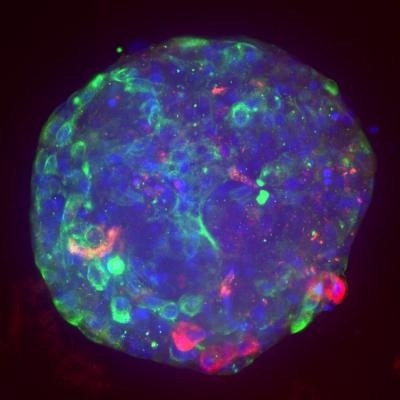
Image Credit: Visikol Inc.
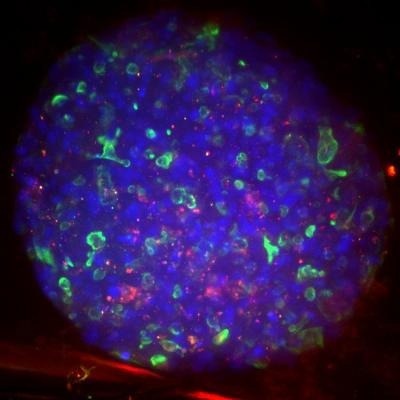
Image Credit: Visikol Inc.
Visikol HISTO-M, a tissue clearing technique made specifically for 3D cell culture models and plate-based high-throughput processing, was created as a solution to this issue.
Complete 3D cell culture model characterization and more precise drug screening are made possible by the Visikol HISTO-M technique when combined with fluorescent labeling (such as fluorescent protein, immunofluorescence, chemical dyes, etc.) and high content confocal microscopy.
Features
Why use Visikol HISTO-M?
- Although numerous tissue clearing methods have been discussed in the literature, only a small number have been proven effective when used with 3D cell culture models
- The majority of these tissue clearing methods are not made to work with well plates, high-throughput processing, or immunofluorescent labeling (e.g., ScaleS4, CUBIC), and many of them have high toxicity (e.g., ClearT)
- The Visikol HISTO-M tissue clearing method was developed to be quick, simple to use, compatible with plates, compatible with fluorescent proteins, and compatible with immunofluorescence
Benefits
- No special equipment required
- Reversible for follow up 2D H&E/IHC
- Compatible with IF, FP, and other fluorescent labels
- Easy-to-use
- Rapid tissue clearing
- Well plate and automation compatible
Visikol® HISTO-M™ Starter Kit. Image Credit: Visikol Inc.
Visikol® HISTO-M™. Image Credit: Visikol Inc.
Application areas
Completed model characterization
Before and after clearing. Image Credit: Visikol Inc.
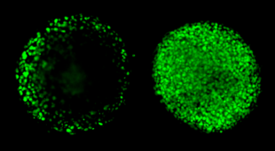
- Through the use of confocal microscopy and Visikol HISTO-M, researchers can fully visualize 3D cell culture models, leading to a 3–4 increase in the number of cells detected
- Both flat bottom plates and U-bottom plates with an extremely low attachment point can be used with this technique
- The comparison between a 3D cell culture model imaged in PBS without clearing (A) and a 3D cell culture model imaged in Visikol HISTO-M (B) demonstrates the benefits of tissue clearing combined with confocal microscopy
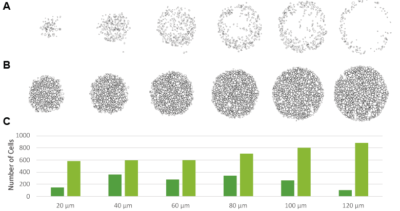
Image Credit: Visikol Inc.
Increased dose response sensitivity
- Due to Visikol HISTO-M’s ability to characterize the entire population of cells within a 3D cell culture model, the technique significantly improves dose response sensitivity
- The evaluation of drug effect is more accurate since the entire cell population is interrogated. Without clearing, only the outer layers of the cells are surveyed, and as a result, measurements of cell proliferation are inflated since the outer layers of the cells exhibit increased proliferation than the inner layers
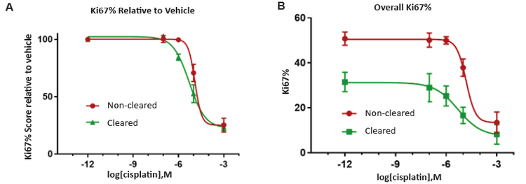
Image Credit: Visikol Inc.