Researchers at the Icahn School of Medicine at Mount Sinai have produced a high-resolution structural map of AAPP (alpha-aminoadipic semialdehyde synthase), an enzyme in the lysine breakdown pathway that they say could be targeted with drugs to help treat glutaric aciduria type 1 (GA1), a rare genetic disorder that can cause brain damage. The study, published in Science Advances, adds details on how the enzyme operates and how its activity is tuned.
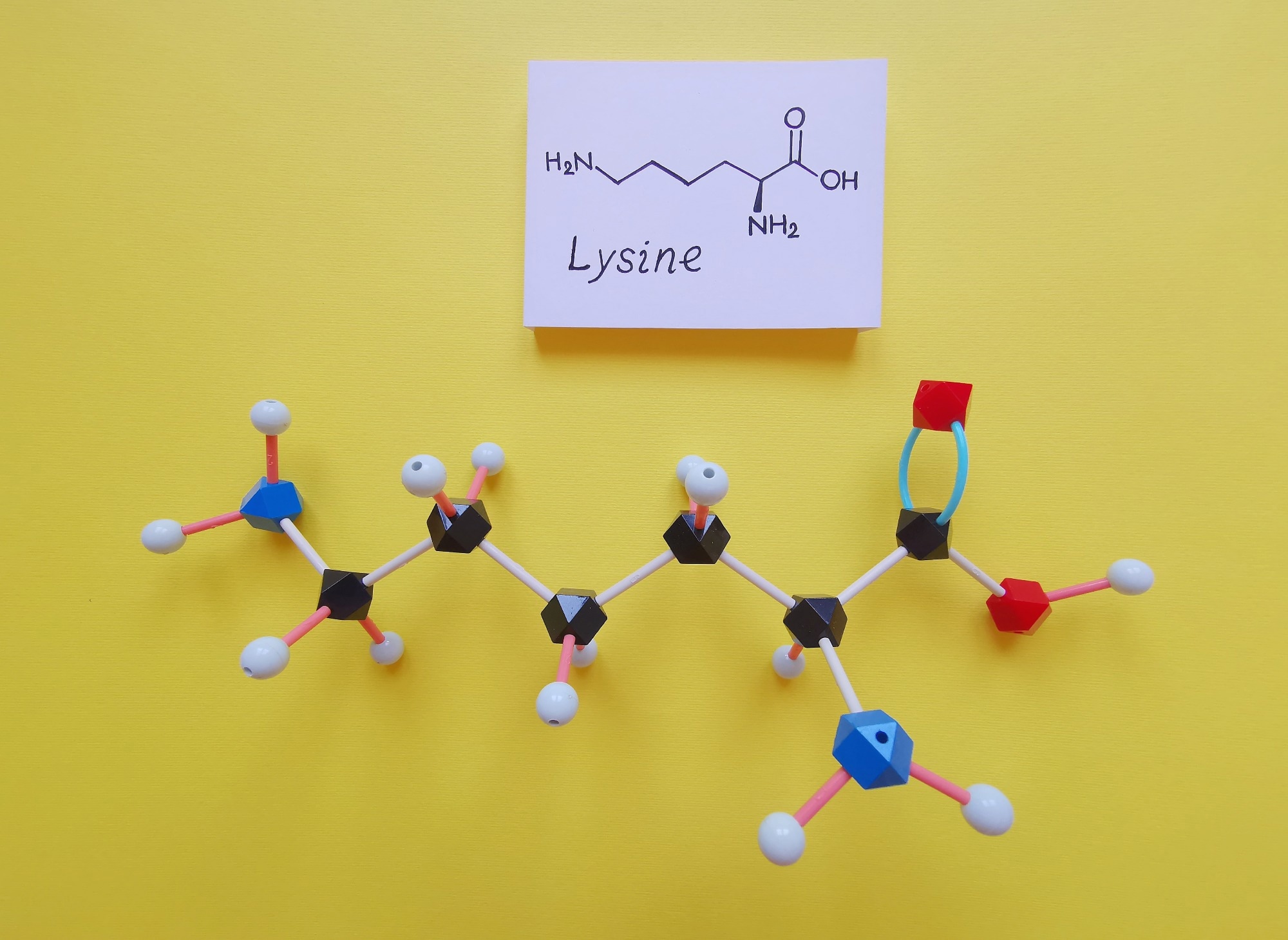 Image credit: Danijela Maksimovic/Shutterstock.com
AASS helps process lysine, and the Mount Sinai team reports identifying a portion of the protein that acts as a regulator of enzyme activity. By clarifying how that control region influences AASS function, the study offers structural guidance for efforts aimed at developing inhibitors, with the long-term goal of limiting the buildup of toxic metabolites linked to GA1.
This is the first detailed picture of the complete AASS enzyme, giving us a much clearer understanding of how its different parts work together. We also found that relatively small changes in a flexible region of the protein can have a substantial effect on its activity. That gives us an important new avenue to explore as we work toward potential treatments for GA1.
Michael Lazarus, PhD, Senior and Study Co-Corresponding Author, Associate Professor, Pharmacological Sciences, and Associate Director, Mount Sinai Center for Therapeutics Discovery, Icahn School of Medicine
GA1 is an inherited metabolic disorder that causes toxic compounds to build up in the body, leading to irreversible brain damage and severe neurological complications.
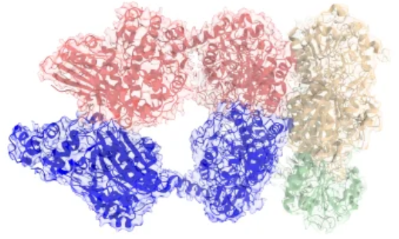 Structure of the full AASS protein determined by cryo-electron microscopy. Image Credit: Wu, Sci.Adv. 12, eaej2268 (2026)
Our investigation of AASS as a potential drug target has revealed multiple ways to alter the enzyme’s activity. A better understanding of how nature uses these mechanisms to control lysine degradation will help with the development of treatments for the disease.
Sander Houten, PhD, Senior and Study Co-Corresponding Author, Professor, Genetics and Genomic Sciences, Icahn School of Medicine
The team first devised a way to isolate the intact human enzyme, then used cryo-electron microscopy to capture high-resolution images of its molecular structure to examine AASS up close. Next, they ran biochemical tests to see how the enzyme behaves under various conditions, including when exposed to activating or inhibiting compounds, while computer simulations showed how shifts in its shape directly affect its activity.
The results highlight a crucial hinge-like region where even subtle structural tweaks trigger major shifts in enzyme function. According to the investigators, mapping this mechanism could pave the way for designing far more effective AASS-blocking drugs.
“Our findings show how a relatively small change at a flexible connection point can influence the behavior of a much larger protein,” said Dr. Lazarus.
With this new enzyme structural knowledge, we can begin to explore how to design molecules that inhibit AASS and assess whether they could eventually lead to a treatment for GA1.
Robert J. DeVita, PhD, Senior and Study Co-Corresponding Author, Professor, Pharmacological Sciences and Director of Medicinal Chemistry, Marie-Josée and Henry R. Kravis Drug Discovery Institute, Icahn School of Medicine, Mount Sinai
The researchers emphasize that these findings are an early step toward a potential therapy rather than an immediate treatment or cure for GA1. Because the work was conducted strictly in the lab, future animal and human studies will be necessary to confirm whether targeting AASS is both safe and effective.
The team plans to use their structural map to screen for and design molecules capable of blocking the enzyme. Beyond advancing GA1 research, the study sheds broader light on how subtle structural shifts can control enzyme behavior.
Source:
Journal reference:
WU, R., et al. (2026) Structural and biochemical insight into allosteric regulation of the human 2-aminoadipic semialdehyde synthase. Science Advances. DOI:10.1126/sciadv.aej2268. https://www.science.org/doi/10.1126/sciadv.aej2268.
 Astrocytic Signaling Pathway Sustains Neuronal Function and Blood Flow