Scientists tracked fluid movement through the human brain and found evidence that cerebrospinal fluid may circulate through brain tissue in a recycling loop, offering new insight into how the brain could clear waste linked to aging and neurodegenerative disease.
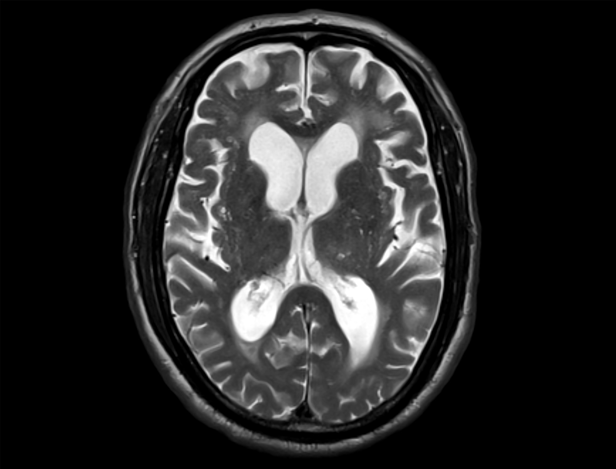 Study: Quantitative assessment of flow between cerebrospinal and interstitial fluid compartments in humans. Image credit: MD Habibur Rahman anis/Shutterstock.com
A recent study published in Proceedings of the National Academy of Sciences suggests that the human brain may use a recirculating fluid loop to clear waste across brain tissues.
Analyzing magnetic resonance imaging (MRI) images using deep-learning-based image analysis tools and mathematical modeling, researchers found that cerebrospinal fluid (CSF) enters brain tissue, exchanges with interstitial fluid (ISF) inside the brain, and then a substantial portion of the fluid appears to return to the CSF around the brain. This suggests that the brain is not a ‘static’ organ but a dynamic system that constantly circulates and recycles fluid in ways that may contribute to waste clearance.
Brain Fluid Recycling May Support Waste Clearance
Scientists suggest that the brain removes waste by moving ISF between brain cells and CSF surrounding the brain and spinal cord. The glymphatic theory suggests that CSF can flow along tiny spaces around blood vessels and enter brain tissue to help clear waste. Animal studies and brain scans in humans support this idea. However, the amount of fluid moving through the brain and the route by which fluid leaves brain tissue have also remained unclear.
MRI Contrast Tracking Reveals Brain Fluid Movement
In the present study, researchers tracked fluid movement in the brain using MRI scans with a gadolinium (Gd)-based contrast agent.
The study included people with suspected idiopathic normal-pressure hydrocephalus (iNPH) and healthy adults aged 65-84 years for comparison. There were 14 patients in the iNPH group and 55 healthy participants. In the iNPH group, the team injected the contrast agent into the fluid around the spinal cord through the intrathecal route. The healthy adults received intravenous injections.
After injecting the contrast agent, the team took MRI scans at different time points. For the intrathecal iNPH group, scans were performed at approximately three hours, five hours, seven hours, and 22 hours after injection. For the healthy adults receiving intravenous injections, scans were obtained at approximately 0.5, 2.7, and 3.2 hours post-injection. Researchers used mathematical models to estimate the amount of CSF entering the brain tissues (CSF-to-ISF inflow) and whether the fluid returned to the surrounding CSF from the brain tissues (ISF-to-CSF recirculation).
Participants maintained the supine position for several hours after injection to minimize movement artifacts on fluid-flow measurements. No serious adverse effects were observed during the study. The team obtained the MRI images using a 3.0 Tesla machine with T1 mapping. They also provided dynamic contrast-enhanced images for the intravenous experiments. These images were analyzed using deep learning and mathematical models. The analysis helped researchers understand fluid exchange in the brain.
MRI Data Suggest CSF Recycling Pathway
The estimated CSF-ISF exchange was larger than previously assumed, and the findings suggest it may play a crucial role in removing waste from the brain. In the iNPH group, nearly 45 mL of CSF moved into the brain tissues every hour. This was surprisingly high since the choroid plexus tissue is believed to produce only about 27 mL of new CSF per hour. The findings suggest that CSF enters the brain tissue before returning to the surrounding fluid, forming a loop rather than flowing unidirectionally.
Most of the fluid exchange occurred in the cortical gray matter, which accounted for approximately 34 ± 14 mL/h of the estimated flow, compared with 11 ± 6 mL/h in white matter and only 0.4 ± 0.3 mL/h in subcortical gray matter. The MRI images also showed that the Gd contrast agent moved through adjacent CSF and brain tissue in similar patterns. The signal, however, appeared in brain tissue slightly later. This delay reinforces the concept of fluid recirculation in waste clearance in the brain. Even when researchers tried different mathematical assumptions, the results were similar, suggesting that the findings are reliable.
Observing the last scan, i.e., 22 hours post-injection, the researchers found that the craniospinal system was not completely clear of the contrast agent. In fact, nearly 36% remained in the brain and spinal cord. Based on this, they estimated a craniospinal outflow rate of about 34–40 mL per hour. The team expected that if CSF moved into brain tissues, then a similar amount of fluid would leave the brain and spinal cord through a distinct drainage pathway.
The measurements, however, did not show such a relationship. After entering the brain tissues, CSF appeared to exchange with ISF to potentially facilitate waste transport, then flow back into the surrounding CSF. Using a continuity-equation approach, the researchers estimated that at least 40% of ISF outflow likely recirculated back into CSF, although the exact proportion remains uncertain. The authors further proposed that this local CSF–ISF recycling system may operate somewhat independently from the separate processes that ultimately remove fluid and waste from the craniospinal system.
The researchers observed similar fluid movement among healthy older adults. After intravenous injections, the contrast showed delayed movement of contrast from the brain tissue into the CSF. Estimated ISF-to-CSF flow rates of 29 ± 74 mL per hour in the full healthy cohort and 58 ± 36 mL per hour in a higher-signal subgroup also supported fluid recycling between brain tissue and CSF. However, the researchers noted substantial variability in these measurements and cautioned that some contrast may have entered CSF directly from blood vessels, potentially leading to overestimation of ISF-to-CSF flow.
Fluid Recycling May Influence Neurodegenerative Disease Risk
The findings suggest that the brain may clear waste through a recycling loop-like fluid system rather than a simple one-way drainage pathway. The results indicate that CSF moves into brain tissue, where it exchanges with ISF, and then returns to the surrounding CSF to help remove waste.
However, the authors emphasized that these volumetric flow estimates depend on the assumption that bulk fluid flow dominates transport. If diffusion plays a larger role, the measurements may instead reflect tracer-specific exchange properties rather than true directional fluid flow.
Further research could aim to improve understanding of the fluid recirculating networks to develop strategies targeting toxic protein buildup in brain tissues in neurodegenerative diseases such as Alzheimer’s disease. Future studies should also include younger and healthier populations and explore the influence of body posture and blood-brain barrier (BBB) leakage on CSF-ISF exchange.
Journal Reference
Wåhlin, A. et al. (2026). Quantitative assessment of flow between cerebrospinal and interstitial fluid compartments in humans. Proceedings of the National Academy of Sciences, 123(18), e2526239123. DOI: 10.1073/pnas.2526239123. https://www.pnas.org/doi/10.1073/pnas.2526239123
 Neuroscientists Identify Brain Cells Acting as the "Disappointment Meter"