Influenza A virus infects hundreds of millions of people each year, yet many of the molecular mechanisms it uses to hijack the machinery inside human cells remain unknown. A team of researchers from Germany applied a chemical labeling and mass spectrometry technique directly inside infected lung cells to develop a detailed map of how viral proteins physically contact human proteins, revealing a previously unrecognized attack on a nuclear structure called the paraspeckle.
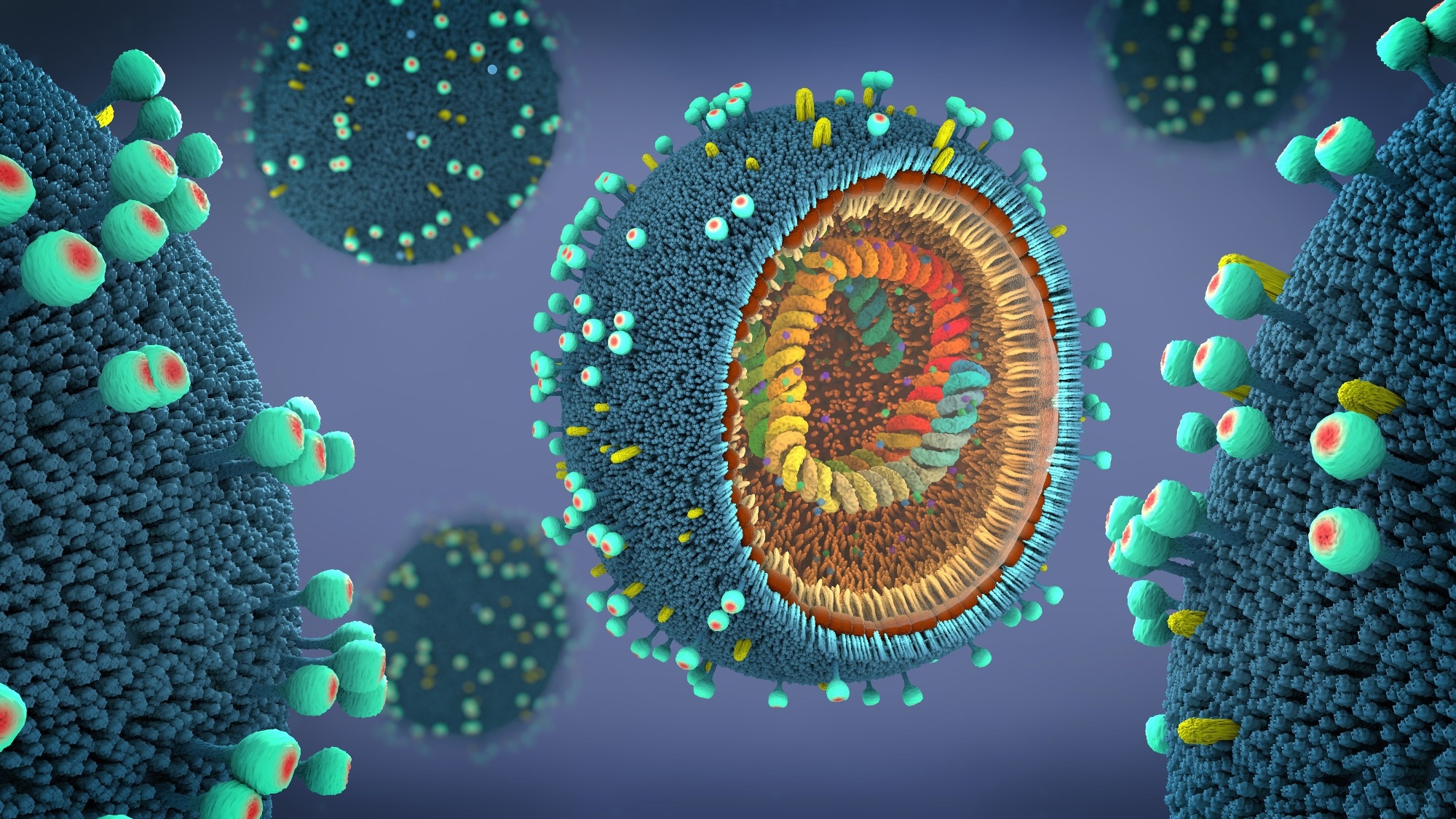 Study: Mapping in-cell protein contact sites reveals hijacking of paraspeckles during influenza A virus infection. Image Credit: Christoph Burgstedt/Shutterstock.com
Background
Influenza A viruses utilize the host cell machinery for nearly every step of their replication cycle, from genome copying to the assembly and release of new virus particles. Although viral proteins interact with human proteins, often within specific cellular compartments and organelles, these host-virus contacts are difficult to capture accurately.
Traditional methods for mapping such interactions typically rely on cells that have been isolated or lysed, which disrupts native architecture and is uninformative about the interactions between viral and human proteins. Although paraspeckles, which are nuclear structures in the host cells built around a long non-coding ribonucleic acid (RNA), are known to influence gene expression and immune signaling, their role during viral infection remains poorly defined.
Want to save this article for later? Click here.
About the study
Researchers applied an in-cell cross-linking mass spectrometry approach known as structural host-virus interactome profiling to human lung epithelial cells infected with the WSN strain of influenza A virus.
They used a modified amino acid to selectively label newly synthesized viral proteins during the later stages of infection, which allowed them to enrich the viral material even though it made up only a small fraction of total cellular protein.
At 14 hours after infection, the cells were treated with a chemical cross-linker that captured proteins in close physical proximity to one another, and the resulting cross-linked peptides were purified, digested with enzymes, and analyzed by mass spectrometry to identify pairs of interacting residues.
The team combined this cross-linking data with computational structural modeling, using AlphaFold-based tools to build three-dimensional models of viral-host protein pairs and to check whether identified contact points were consistent with plausible molecular interfaces.
The selected interactions were independently validated using reciprocal immunoprecipitation combined with mass spectrometry and proximity ligation assays, which visualize when two proteins are in proximity to each other inside intact cells.
To test which host factors mattered functionally, the researchers performed a short-interfering RNA knockdown screen targeting the proteins identified in the cross-linking network. They then measured the effects of those proteins on viral replication using a luciferase reporter system and standard viral titration assays.
The researchers also used clustered regularly interspaced short palindromic repeats (CRISPR)-associated protein 9 (Cas9) gene editing to entirely delete several host genes to generate knockout cell lines.
The team used fluorescence in situ hybridization (FISH) to visualize and quantify nuclear RNA-protein structures across several infected cell types, including primary human airway cells.
Key findings
The results revealed that influenza A viruses make extensive physical contact with human proteins inside infected cells, forming hundreds of distinct interactions that trace out specific biological pathways rather than occurring at random.
Many of these contacts clustered around the maturation of the viral surface protein hemagglutinin as it moved through the endoplasmic reticulum and Golgi apparatus. Moreover, knockdown of several newly identified host factors along this route reduced viral infection, confirming their functional relevance to viral protein processing.
Interestingly, the study found that two viral proteins interacted extensively with paraspeckle components, and infection consistently triggered a progressive loss of paraspeckle structures within the first several hours. This effect was observed across multiple cell types, including primary airway cells, and across several different influenza strains.
The researchers proposed that this disassembly of paraspeckles happens through three combined mechanisms: direct viral protein interactions with paraspeckle proteins, degradation of the structural RNA by a viral enzyme, and interference with the host cell's transcription machinery.
Removing individual paraspeckle proteins through gene knockout increased viral replication. The researchers proposed that their disassembly frees up host RNA-binding proteins that the virus can then recruit to support its own replication, while potentially also weakening antiviral gene responses.
The findings suggested that individual influenza strains may rely on different components of the pool of released host proteins.
However, the authors noted a key limitation: the cross-linking approach favors proteins present in sufficient abundance and does not capture very early or highly transient interactions, suggesting that some relevant host factors, particularly at earlier infection stages, might have gone undetected in the study.
Conclusions
Overall, the study provided a spatially detailed map of how the influenza A virus interacts with human proteins inside living cells, which uncovered coordinated viral strategies that spanned surface protein maturation and nuclear organization.
The researchers also linked specific molecular contacts to functional outcomes and identified new host factors involved in haemagglutinin processing and paraspeckle disruption. These findings offer promising potential targets for future antiviral strategies.
Journal reference:
Kotova, I., Mühlberg, L., Gilep, K., Burtscher, M. L., Becher, I., Yu, D., Ziemianowicz, D., Stanelle-Bertram, S., Beck, S., Baeg, K., Grba, A., Duss, O., Gabriel, G., Savitski, M. M., Liu, F., Bogdanow, B., & Kosinski, J. (2026). Mapping in-cell protein contact sites reveals hijacking of paraspeckles during influenza A virus infection. Nature Microbiology. DOI:10.1038/s41564-026-02416-1, https://www.nature.com/articles/s41564-026-02416-1
 Groundbreaking ARUP Research Clarifies Clinical Value of Atypical Cell-Free DNA Screening Results