Cells subjected to intense, sustained mechanical strain must protect the nucleus, which is the most vital structure. Keratin filaments are best known for reinforcing cellular components against damage. However, a recent study published in Nature Physics revealed that when cells are stretched for hours, these filaments undergo a dramatic structural transformation, bundling into thick radial spokes and ultimately releasing the cell's nucleus from its protective mesh.
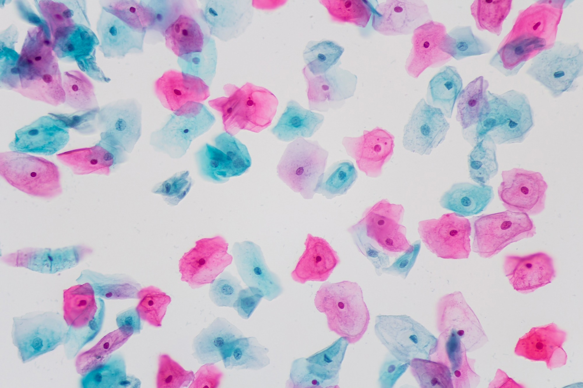 Study: Dynamics of supracellular keratin bundling and nuclear uncaging in stretched epithelia. Image Credit: Komsan Loonprom/Shutterstock.com
Background
Cells and tissues constantly encounter mechanical forces, from the routine expansion of the bladder to the large deformations experienced during development. A family of cytoskeletal polymers known as intermediate filaments, which includes keratin, is considered central to how cells withstand this stress.
In the relaxed state, keratin forms a connected network, with one part lining the cell surface and another enclosing the nucleus. The two parts are linked by short filament bridges. This arrangement, described as a rim-and-spoke structure, depends on crosslinking proteins that tie keratin to the actin cytoskeleton and to the nuclear envelope.
Despite broad agreement that intermediate filaments guard cells against large-scale deformation, exactly how their architecture changes during sustained stretching, and how the change impacts the nucleus, has remained unclear.
Want to save this article for later? Click here.
The Study
Researchers have used canine kidney cells engineered to express fluorescently tagged keratin-18, along with markers for the plasma membrane and nucleus, to visualize cytoskeletal architecture in living epithelial monolayers.
The team used two complementary systems to generate a controlled mechanical strain. In one system, micropatterned spontaneous domes formed naturally in monolayers due to osmotic pressure, while in the other, a custom-built microfluidic device applied defined hydrostatic pressure beneath a porous membrane, producing domes of consistent size with adjustable inflation over time.
Confocal time-lapse microscopy was used to track keratin organization in these domes for several hours, and cells were classified according to two quantitative metrics. The intensity radius described whether keratin accumulated centrally or peripherally within a cell, while the average angle described whether the filaments were arranged radially or circumferentially.
To probe the role of the actin cytoskeleton, the researchers disrupted plectin, the principal protein linking keratin to actin, by overexpressing a truncated, dominant-negative version that retains actin binding but loses its keratin-binding capacity.
The team also used a separate set of experiments with the drug latrunculin A to rapidly dismantle actin filaments altogether. A deoxyribonucleic acid (DNA)-binding fluorescent dye was used to simultaneously visualize keratin and nuclear signals, which allowed the team to measure the physical offset between the keratin network and the nucleus over time.
Moreover, to interpret these observations mechanistically, the researchers built computational models using a customized version of the cytoskeletal simulation platform Cytosim, first in a simplified two-and-a-half-dimensional geometry and later in a full three-dimensional format incorporating a mobile nucleus.
Finally, to determine whether the bundling of keratin fibers spread through tissues randomly or through a directed process, the team employed time-lapse imaging of multiple domes with simulations and compared unbiased and neighbor-dependent probabilities of individual cells adopting the bundled state.
Sustained Mechanical Stretching Reorganizes Keratin Cytoskeleton
The study found that sustained mechanical stretching resulted in a slow but dramatic reorganization of the keratin cytoskeleton, ultimately causing the cell nucleus to lose its protective covering entirely.
Rather than responding immediately to stretching, the keratin filaments depleted from tricellular junctions only after several minutes, gradually consolidating into thick bundles that radiated outward from a central point, a configuration the researchers termed a star-like arrangement.
This transition to a star-like arrangement took an average of roughly seven hours to complete and, once established, did not reverse even after the tissue relaxed and reinflated. This indicated that the change was effectively permanent within the timeframe studied.
The reorganization also proved highly dependent on interactions between keratin and the actin cytoskeleton. Disruption of the crosslinking protein plectin nearly tripled the speed of bundling, while chemically dissolving actin filaments using latrunculin A triggered comparably rapid keratin reorganization.
Computational modeling results supported these observations and revealed the underlying physical mechanism. It showed that stretching initially tightened keratin around the nucleus, but this tension ultimately generated a compressive force strong enough to squeeze the nucleus free of its keratin cage.
Live imaging confirmed this prediction, showing a progressive and eventually complete separation between keratin and nuclear signals as the bundling process advanced. At the tissue level, bundling did not occur randomly across cells but instead spread through a nucleation-and-growth process, with cells showing a greater probability to bundle if a neighboring cell had already done so. This clustering behavior, verified through simulations, also suggests that mechanical or biochemical cues pass between neighboring cells during the transition.
The authors noted that whether losing the protective keratin cage ultimately helps or harms the nucleus remains unresolved. While an exposed nucleus may be more vulnerable to mechanical damage, its separation from a highly stressed cytoskeleton could also offer a different form of protection against force transmission.
Conclusion
The study identified a previously undescribed structural transition in the keratin cytoskeleton, where they undergo a dramatic transition in response to prolonged mechanical stress.
The findings raise new questions about cellular resilience during development, organ stretching, and disease, while also offering a physical framework for future studies of tissue mechanics and bioinspired materials.
Journal reference:
Golde, T., Pensalfini, M., Chahare, N., Roca-Cusachs, P., Wiche, G., Charras, G. T., Arroyo, M., & Trepat, X. (2026). Dynamics of supracellular keratin bundling and nuclear uncaging in stretched epithelia. Nature Physics. DOI:10.1038/s41567-026-03371-8, https://www.nature.com/articles/s41567-026-03371-8
 Groundbreaking ARUP Research Clarifies Clinical Value of Atypical Cell-Free DNA Screening Results