Doctors have long attributed the persistent and widespread pain reported by colorectal cancer (CRC) survivors to painful nerve damage from surgery or chemotherapy. However, a recent study published in Nature Communications suggested that the cancer itself may be causing nerve damage. Researchers found that colorectal tumors triggered inflammatory nerve damage in both mice and primates.
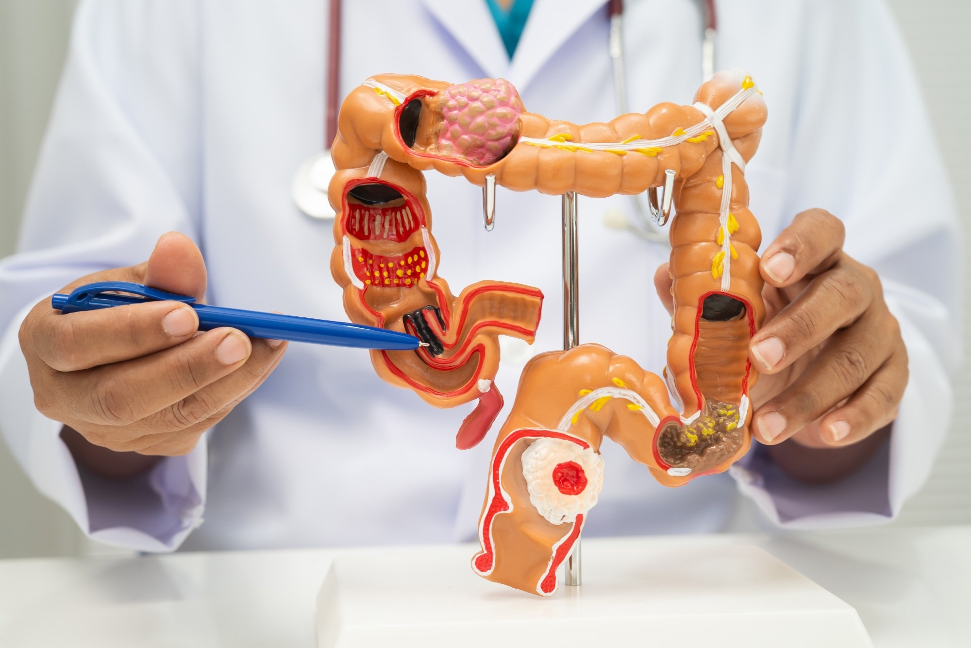 Study: Inflammatory neuropathy in mouse and primate models of colorectal cancer. Image Credit: sasirin pamai/Shutterstock.com
Background
Improved tumor detection and treatment have boosted CRC survival, and medical focus has now shifted to addressing lasting complications such as chronic pain and neuropathy, which affect many survivors. Surgery and chemotherapy are typically blamed for nerve damage, but treatments for chemotherapy-induced neuropathy remain limited. Moreover, the underlying cause of the lasting pain in some survivors also remains poorly understood.
While prior nervous system injury is a known risk factor, the cancer’s direct role is rarely considered outside rare paraneoplastic syndromes tied to lymphoma and lung cancer, where tumors trigger antibodies against neuronal tissue. Whether common cancers such as CRC independently damage peripheral nerves has hitherto remained largely unexplored.
Want to save for later? Click here.
About the Study
Researchers at MD Anderson Cancer Center at the University of Texas used an orthotopic mouse model of CRC to investigate whether tumor growth alone could damage peripheral nerves.
They used colon cancer cells representing microsatellite-instability-high disease (MC38), which were injected directly into the rectal submucosa of mice under anesthesia. The researchers tracked tumor growth weekly using bioluminescent imaging. Most experiments were conducted three weeks after injection, which is the point at which neuropathy had previously been shown to emerge in this model.
To assess sensory and motor function, the team applied numerous behavioral tests, including von Frey filament testing for tactile sensitivity, a thermal gradient ring for temperature preference, a burrowing assay for pain-related behavior, and automated gait analysis using video-tracking equipment.
Mice also completed a beam-walking task across beams of increasing difficulty to measure motor coordination. A subset of tumor-bearing mice also received the anti-inflammatory drug carprofen in their drinking water to determine whether inflammation contributed to any behavioral changes observed.
Skin biopsies were immunostained to measure nerve fiber density. The sciatic nerve and dorsal root ganglia tissue were examined using immunohistochemistry, transmission electron microscopy, and bulk ribonucleic acid (RNA) sequencing to detect signs of Schwann cell injury, demyelination, and immune cell infiltration.
The researchers characterized plasma and nerve lipid profiles through untargeted lipidomics using liquid chromatography-mass spectrometry.
To probe the specific role of macrophages, they used a genetic mouse model that allows chemogenetic depletion of these immune cells through targeted drug administration. The team evaluated calcium signaling and electrical activity in sensory neurons through live-cell imaging and multi-electrode array recordings of cultured ganglia.
Lastly, to test whether these findings extended beyond mice, the team analyzed archived plasma and sciatic nerve tissue from a colony of rhesus macaques that spontaneously develop microsatellite-instability-high CRC, and compared the animals with confirmed cancer diagnoses at necropsy to cancer-free controls of similar age.
Key Findings
The study found that colorectal tumor growth triggered inflammatory nerve damage in mice, independent of chemotherapy or surgery, and that comparable changes also occurred naturally in primates with the disease. Mice with tumors showed reduced nerve fiber density in the skin without clear increases in tactile or thermal sensitivity. They also showed subtle decreases in gait and motor coordination that were partly reversed by the anti-inflammatory drug carprofen, suggesting inflammation as an early driver of these changes.
The researchers observed that tumor growth was accompanied by widespread disruption of plasma lipid composition and substantial loss of triglycerides, along with increased levels of inflammatory lipid classes such as sphingolipids and ceramides. Similar effects were observed in the sciatic nerve itself, along with signs of Schwann cell injury, such as the decompaction of the myelin sheath surrounding nerve fibers. Electron microscopy also confirmed structural changes across axons of multiple sizes, consistent with impaired nerve conduction.
The study also found that immune involvement extended beyond generalized inflammation. The sensory ganglia showed antibody deposit accumulation resembling patterns seen in classic paraneoplastic neuropathies linked to other cancers, and this response was absent in mice lacking mature antibody-producing immune cells.
Macrophages also accumulated in both nerves and ganglia and shifted toward a pro-inflammatory profile. However, depleting macrophages appeared to worsen motor performance, suggesting these cells play a partly protective role despite contributing to inflammation.
Rhesus macaques that developed CRC independently showed comparable plasma lipid disruption, elevated inflammatory markers, and clear nerve inflammation and degeneration compared to cancer-free animals. However, the authors noted that separating the individual contributions of inflammatory signaling from lipid dysregulation remains difficult, since many inflammatory mediators are themselves lipid-derived.
Conclusion
Overall, the findings establish a link between CRC and inflammatory nerve damage, driven by a combination of dyslipidemia, immune cell infiltration, and antibody deposition that disrupts sensory neuron and Schwann cell function.
Because this damage appeared consistently across mice and primates, the authors suggested CRC may represent an underappreciated, pre-existing risk factor for neurological complications in survivors, potentially compounding nerve injury from later chemotherapy exposure. However, further research is needed to identify the specific antibody targets involved.
Journal reference:
Gaffney, C. M., Casaril, A. M., Mahmud, I., Wei, B., Jiang, Y., Fiore, N. T., Liu, J., Valadez, K. M., Kolb, E. A., Cherry, F. R., Shi, L., Dunner, K., Lorenzi, P. L., Reiken, S. R., Grace, P. M., Mahalingam, R., Guise, T. A., Hodo, C. L., & Shepherd, A. J. (2026). Inflammatory neuropathy in mouse and primate models of colorectal cancer. Nature Communications. DOI:10.1038/s41467-026-76683-1
https://www.nature.com/articles/s41467-026-76683-1
 Higher-Order Receptor Clustering Acts as Hidden Lymphangiogenic Signal Amplifier