The adult human heart ventricle contains various supporting cell types, such as endothelial cells, cardiomyocytes, and fibroblasts. Although cardiomyocytes account for 75% of the total volume of the human ventricle, they only make up 50% of the total cell number.
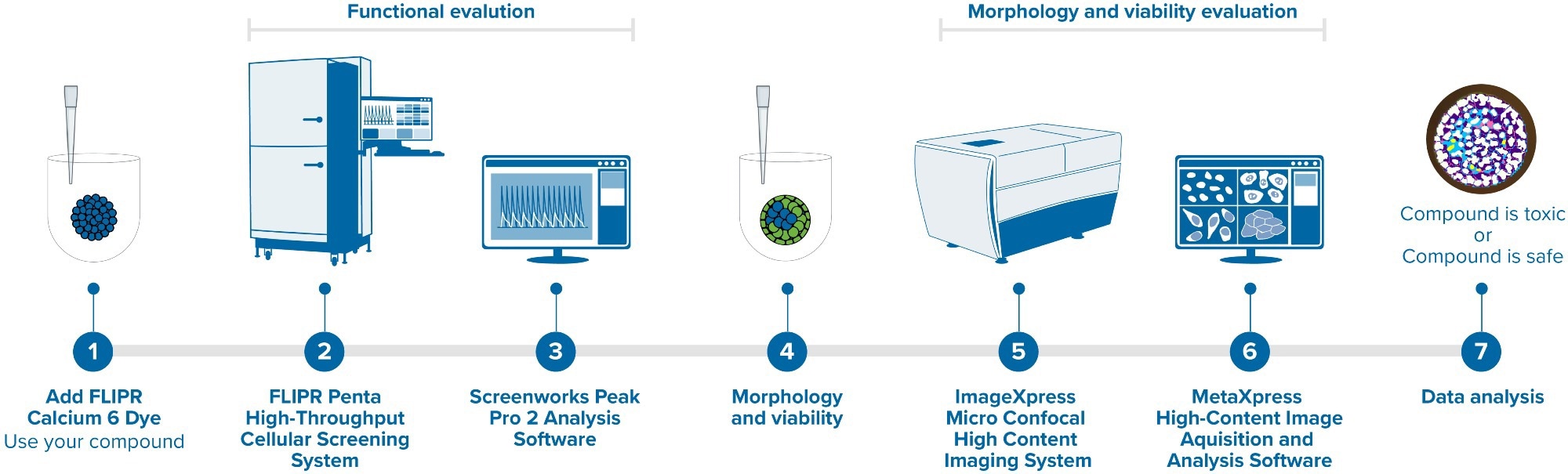
Image Credit: Molecular Devices UK Ltd
In recent publications, the tri-cellular co-culture microtissues of endothelial cells, cardiomyocytes, and cardiac fibroblasts (all derived from human iPSC) have been shown to improve the maturation and functional activity of cells in comparison to 2D cardiomyocytes.
This means that the tri-cellular co-culture microtissues are more representative of the real physiology of the human heart.
This article presents a study using a tri-culture model generated by mixing iPSC-derived cardiac and endothelial cells with primary adult fibroblasts in a ratio of 75:15:10 in ultra-low attachment (ULA) plates directly from thaw.
Benefits
- The tri-culture model provides a novel tool for the evaluation of functional and morphological effects on cardiac cells.
- The FLIPR Penta System allows better resolution of calcium oscillation patterns in cardiomyocytes.
- MetaXpress CME allows individual cells to be located and 3D structures of cell models to be reconstructed.
A Biomek i7 Automated Workstation, acquired from Beckman Coulter Life Sciences, was utilized for cell plating and the following media exchange every two days. The formation of 3D microtissues took place within 48 hours. These microtissues then began to spontaneously and regularly contract on the fifth day.
The functional activity of microtissues was assessed through readings of calcium oscillations after a calcium dye was added. These readings were taken using a fast kinetic fluorescence recording instrument, the FLIPR® Penta High-Throughput Cellular Screening System, as presented in Figure 1.
The microtissues’ response to various known modulators of cardiac activity was tested. Results showed that molecules like isoproterenol substantially accelerated the oscillation rate as well as increased the peak amplitude (inotropic response).
The changes in the Ca2+ oscillation patterns from other compounds, including hERG inhibitors, ion channel blockers, or beta-blockers, were consistent with the expected mode of action. Waveform analysis was carried out with the ScreenWorks® Peak Pro 2™ software module.
For the characterization of the structure and morphology of the 3D microtissues, the ImageXpress® Micro Confocal High-Content Imaging System is utilized for high-content imaging, as shown in Figure 1.

Figure 1. FLIPR Penta High-Throughput Cellular Screening System and ImageXpress Micro Confocal High-Content Imaging System. Image Credit: Molecular Devices UK Ltd
The various cell types were immunostained with antibodies specific for VE-Cadherin (for endothelial cells), Troponin T (for cardiomyocytes), and COL1A1 (for fibroblasts). The 3D structure of the microtissues was reconstructed and subsequently analyzed with the MetaXpress® High-Content Image Acquisition and Analysis Software.
The results of this study demonstrate the efficiency and biological relevance of iPSC-derived cell types in 3D micro-tissues as a promising tool for measuring the compound effects on human cardiac tissues in a high-throughput format.
Instruments
This experiment utilized a high-speed EMCCD camera on the FLIPR Penta High-Throughput Cellular Screening System to take measurements of the patterns and frequencies of the Ca2+ oscillations of cardiac tri-culture microtissues.
These were monitored by changes in the intracellular Ca2+ levels using the EarlyTox™ Cardiotoxicity Kit, acquired from Molecular Devices.
The instrument was used in combination with the ScreenWorks PeakPro2 peak analysis software to conduct analysis and characterization of the primary and secondary peaks and complex oscillation patterns.
The ImageXpress Micro Confocal HighContent Imaging System was utilized in combination with an sCMOS camera and a spinning disk confocal to enable the 3D structures of the whole microtissues to be obtained. These microtissues were stained with antibodies in different fluorescent channels.
For the reconstruction of the 3D structure of the microtissues, the custom module editor was employed, which may be customized for sufficient image analysis.
Methods
Cryopreserved human iPSC-derived iCell® Cardiomyocytes2 and iCell Endothelial Cells from FUJIFILM Cellular Dynamics, Inc. were utilized, as well as Primary Human Cardiac Fibroblasts Cells from Promocell. These were then seeded at a 75:15:10 ratio. The time point of the workflow is detailed in Figure 2.
Strong synchronous contractions were shown to be present in the 3D cultures via visual confirmation before the experiments were run.
The cells were stained to enable the study of phenotypic changes using a mixture of the Hoechst nuclear dye (2 μM) and the viability dye Calcein AM (1 μM), both from Life Technologies.
Measurements of the calcium oscillations in the cells were taken using the FLIPR Penta instrument following the compound treatment. They were subsequently fixed and stained with either anti Troponin T A647 and anti COL1A1 A568 or anti Troponin T A647 and anti VE-Cadherin A568.

Figure 2. The workflow of the iPSC-derived Cardiac Tri-Culture Model. Image Credit: Molecular Devices UK Ltd
Results
Kinetic Pattern Recording and Analysis
On the day of the assay, the EarlyTox Cardiotoxicity Kit was utilized to load cardiac tri-culture microtissues. They were then treated with compounds for one hour. A rate of 30–50 frames per second was used to record spontaneous calcium oscillations to enable proper resolution of the complex oscillation patterns.
Advanced analytical methods were employed for the multi-parametric characterization of the Ca2+ flux oscillation patterns. This phenotypic assay enables measurements to be characterized, such as oscillation frequency, amplitude, irregularity, peak rise and decay times, as well as peak width.
In addition, the appearance of early-afterdepolarization (EAD)-like event patterns, peak prolongation, and peak irregularity was evaluated. Representative tracing curves of the fluorescence signals are shown in Figure 3.

Figure 3. The tracing curve of fluorescence signals of Isoproterenol at 0.04, 0.15, and 2.5 μM; E-4031 at 0.04, 0.15, 2.5, and 10 μM; and Chlorpromazine at 0.15, 0.6, 2.5, and 10 μM. Image Credit: Molecular Devices UK Ltd
Parametric Effects of Compounds
A summary of the trend of effects of eight modulators on the cardiac activity of microtissues is presented in Figure 4, where parameters like peak counts/300 seconds, amplitude (RFU), and CTD 90 (seconds) are shown.
As expected, Isoproterenol and other well-recognized antagonists accelerate the beat rate and decrease CTD90. Alternative antagonists, including cisapride and chlorpromazine, slow the beat rate and amplitude after a particular threshold of concentration is met.
A summary of the EC50 values of the compound concentration is detailed in Table 1.
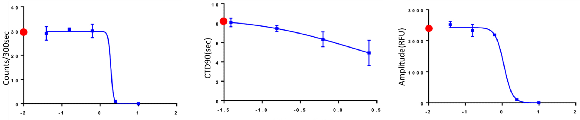
Figure 4. The trend of the change of Peak Counts/300 sec, CTD90 and Amplitude with the increasing concentration of compounds. Red dots indicated control values for appropriate measurements. Image Credit: Molecular Devices UK Ltd
Table 1. Summary chart of EC50 values (µM) calculated with GraphPad Prism. Source: Molecular Devices UK Ltd
| Compound |
Peak Count per 300 sec |
CTD90 (width of peak/sec at 90% of height) |
Amplitude |
MOA |
| Isoproterenol |
1.25 |
0.35 |
12.76* |
B2 adrenoreceptor agonist |
| Cisapride |
1.9* |
1.4 |
1.1 |
hERG blocker, antacid |
*denotes an ambiguous value in Prism.
High-Content Imaging of 3D Cardiac Microtissue Structures
High-content imaging of the microtissues was performed using TRITC, Cy5, and DAPI channels, with several z-planes captured to cover the entire microtissue. The MetaXpress built-in custom module editor was employed to analyze the 3D coordinates of each cell from the resultant images.
Figure 5 shows the representative overlay images of the combination of cardiomyocytes and fibroblast cell staining and the combination of cardiomyocytes and endothelial cell staining.

Figure 5. Composite images of cardiac tri-culture microtissues. After fixation and permeabilization, cells were stained either with a combination of anti-Troponin T AF647 + anti-VE-Cadherin AF568 or anti-Troponin T AF647 + anti-COL1A1 AF568. Cells were imaged with the DAPI, TRITC and Cy5 channels, 10X Plan Fluor objective. (A) Red: iCell Cardiomyocytes, Green: Cardiac Fibroblast Cells, Blue: Hoechst; (B) Red: iCell Cardiomyocytes, Green: iCell Endothelial Cells, Blue: Hoechst; (C) 3D reconstruction of fibroblast cells from the microtissues shown in Figure 5A. Image Credit: Molecular Devices UK Ltd
These images indicate that the cardiac fibroblast cells are more likely to cluster compared to endothelial cells. A silhouette analysis was performed to further examine this phenomenon.
3D Reconstruction of Cell Distribution and Silhouette Analysis of Clustering
A silhouette score was employed to assess the clustering of the cardiac fibroblast cells. These scores ranged from -1 to +1 to indicate how far each point in a cluster is from points in the neighboring clusters, where +1 represents the point being far away from neighboring clusters.
The 3D reconstruction of the fibroblast cells displayed in Figure 5A is presented in Figure 5C, where a total of four clusters can be seen, gaining the highest silhouette score (data not shown).
The overall silhouette analysis of fibroblast cells proves the higher tendency of fibroblasts to produce clusters. However, the impact of this tendency on the maturation of cardiac microtissues requires further investigation.
Conclusions
This study considered tri-culture models that were produced by mixing iPSC-derived iCell Cardiomyocytes2 with iPSC-derived iCell Endothelial Cells and primary adult fibroblasts. This was shown to be a novel tool for assessing the functional and morphological effects on cardiac cells.
The FLIPR Penta System was utilized in combination with a new high-speed camera to allow enhanced resolution of calcium oscillation patterns in cardiomyocytes, while the ImageXpress Micro Confocal high-content imaging system was employed for the rapid and high-resolution acquisition of complex biological samples.
The MetaXpress custom module editor proved to be a powerful analysis tool that enabled the location of the individual cells and the reconstruction of the 3D structure of complex cell models.
A range of parametric responses was shown through the use of a set of eight known modulators of cardiac activities. This assay can be utilized to test developing drugs as well as screen chemicals for potential cardiotoxic hazards.
About Molecular Devices UK Ltd
Molecular Devices is one of the world’s leading providers of high-performance life science technology. We make advanced scientific discovery possible for academia, pharma, and biotech customers with platforms for high-throughput screening, genomic and cellular analysis, colony selection and microplate detection. From cancer to COVID-19, we've contributed to scientific breakthroughs described in over 230,000 peer-reviewed publications.
Over 160,000 of our innovative solutions are incorporated into laboratories worldwide, enabling scientists to improve productivity and effectiveness – ultimately accelerating research and the development of new therapeutics. Molecular Devices is headquartered in Silicon Valley, Calif., with best-in-class teams around the globe. Over 1,000 associates are guided by our diverse leadership team and female president that prioritize a culture of collaboration, engagement, diversity, and inclusion.
To learn more about how Molecular Devices helps fast-track scientific discovery, visit www.moleculardevices.com.
Sponsored Content Policy: AZO Life Sciences publishes articles and related content that may be derived from sources where we have existing commercial relationships, provided such content adds value to the core editorial ethos of News-Medical.Net which is to educate and inform site visitors interested in medical research, science, medical devices and treatments.