In pharmaceutical, environmental, and toxicology labs, live-cell-based assays are routinely conducted to investigate cell growth, viability, cytotoxicity, and numerous other parameters.
When evaluating how cells respond to pharmaceutical or compound treatment, frequent monitoring of cellular characteristics over multiple days is often necessary for determining the optimal treatment window for a specific cell or 3D culture model.
Maintaining consistent cell health with these assays is critical to ensure data reliability and sensitivity. For example, temperature and gas composition are important variables that must be managed to support assay reproducibility. Ideally, maintaining healthy cells requires mimicking incubator conditions inside a microplate reader.
Most modern microplate readers include a temperature-control feature that maintains a constant 37 °C throughout extended cell-based experiments. Furthermore, equipping a reader with a gas mixer capable of supplying a specified mix of gases, including above-ambient CO2, can more closely replicate incubator conditions and support optimal cell health over lengthy durations.
This paper demonstrates the enhancement in cell viability achieved using a SpectraMax® iD5e or iD3s Multi-Mode Microplate Reader with a SpectraMax® aer Gas Mixer compared to a reader lacking a gas mixer. U-2 OS cells were grown in 96-well plates for 72 hours under various incubation conditions.
Cell viability was determined using an endpoint luminescent ATP assay at 72 hours or monitored at intervals throughout the time course with the RealTime-Glo™ MT Cell Viability Assay.
Benefits
- Gas mixer allows precise adjustment of the cell culture environment
- Three-day cell viability comparable to that obtained using a conventional cell culture incubator
- User-friendly interface simplifies gas mixer configuration
Materials
- U-2 OS osteosarcoma cells (ATCC cat. #HTB-96)
- Growth medium for U-2 OS cells:
- McCoy’s 5A (Modified) Medium (Thermo Fisher cat. #16600082)
- 10% fetal bovine serum (FBS, Avantor® Seradigm cat. #1500-500)
- Penicillin/streptomycin (Thermo Fisher Scientific cat. #15070-063)
- RealTime-Glo MT Cell Viability Assay (Promega cat. #G9711)
- CellTiter-Glo 2.0 Cell Viability Assay (Promega cat. #G9241)
- 96-well black-walled, clear-bottom cell culture microplates (Corning cat. #3904 or 3603)
- ibiSeal self-adhesive cover film, 76.0 mm × 114.0 mm, sterilized (ibidi cat. #10874)
Methods
- Cell plating: U-2 OS cells from a single confluent T75 flask were trypsinized and suspended in 20 mLs media before counting. Cell viability was 90%, with 3.3 x 10⁵ live cells/mL.
Following dilution in media, the cells were seeded into wells of 96-well black/clear cell culture microplates at 4000, 2000, and 1000 cells/well, using 100 μL per well and 18 wells for each seeding density. Edge wells were filled with 200 μL/well of PBS, and ibiSeals were applied to reduce evaporation.
- Assay setup: RealTime-Glo MT assay reagents were added to half of the wells containing cells, with a final well volume of 200 μL. These wells were monitored at one-hour intervals throughout the three-day incubation period.
- Incubation conditions: Plate incubation occurred under three different conditions: (1) a cell culture incubator at 37 °C/5% CO2; (2) a SpectraMax iD5e or iD3s Multi-Mode Microplate Reader with SpectraMax aer Gas Mixer, set to 37 °C with 5% CO2 (Figure 1); and (3) a SpectraMax reader configured to 37 °C with ambient CO2 (no gas mixer).
- RealTime-Glo MT assay: Assay plates were read by the luminescence detection mode of the SpectraMax iD5e or SpectraMax iD3s reader. Throughout the three-day incubation inside the reader, luminescence readings were collected each hour. Growth curves were displayed as kinetic traces by SoftMax® Pro Software, while viability results were plotted by taking the maximum minus minimum signal for every well.
- CellTiter-Glo 2.0 assay: Following the three-day incubation, CellTiter-Glo™ 2.0 (Promega) reagent was dispensed into the wells. After shaking for two minutes, the plates were incubated at ambient temperature for 10 minutes. Luminescence was subsequently read on a SpectraMax iD5e or iD3s reader.
- Data analysis and graphing: Data production was supported using preconfigured protocols in SoftMax Pro software. All data was evaluated and results plotted using this software.
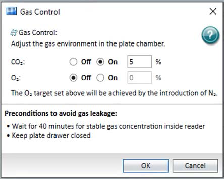
Figure 1. Gas control user interface in SoftMax Pro Software. CO2 levels from 0.1% to 15.0%, and O2 levels from 1.0% to 21.0%, can be set here. Image Credit: Molecular Devices UK Ltd
Results
The growth curves shown in Figure 2 were generated by monitoring growth via luminescence measurements with RealTime-Glo MT at one-hour intervals. Cells seeded at greater densities exhibited a decline in growth rate during the third day of growth, while cells seeded at the lowest density continued to grow at a more constant rate.
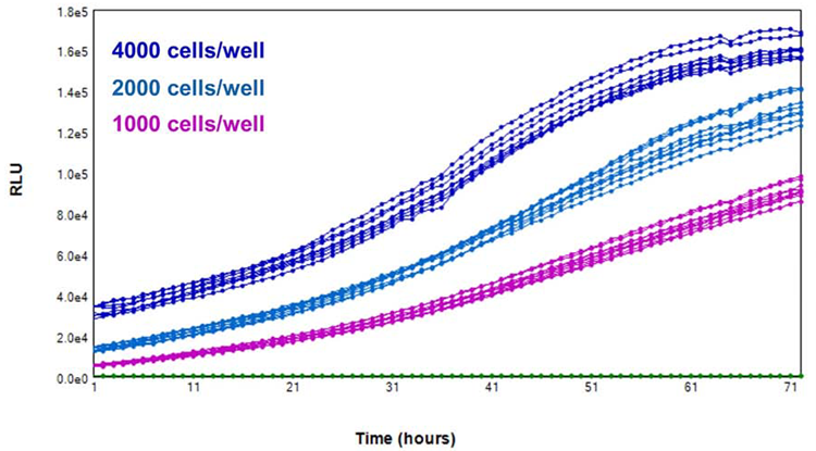
Figure 2. Growth monitored over the course of 3 days inside a SpectraMax iD5e reader with a SpectraMax aer gas mixer, using the RealTime-Glo MT assay. Image Credit: Molecular Devices UK Ltd
As anticipated, cells assayed using the luminescent ATP assay exhibited stronger signals at elevated seeding densities following three days in culture. Compared with endpoint measurements from the RealTime-Glo MT-assayed cells on day three, the overall RLU magnitudes were greater with the ATP assay.
However, the trends between RLU and cells seeded per well were comparable, showing that either assay can be effectively used to evaluate viability (Figure 3).
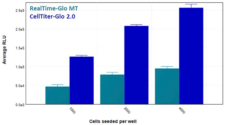
Figure 3. Cell viability after three days in a SpectraMax iD3s reader at 37 °C/5% CO2, with RealTime-Glo (light blue) and CellTiter-Glo (dark blue) readouts. Image Credit: Molecular Devices UK Ltd
Results
Cell viability was better in cells incubated in SpectraMax readers equipped with a gas mixer than in cells incubated in a plate reader with no gas mixer (Figure 4). The SpectraMax aer gas mixer, configured to supply 5% CO2 to the plate reader’s read chamber, enabled cell health more closely resembling that exhibited when cells were maintained in a conventional cell culture incubator.
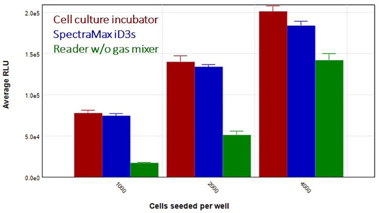
Figure 4. Comparison of cell viability after three days of incubation in a cell culture incubator (red), SpectraMax iD3s reader with SpectraMax aer gas mixer (blue), and a SpectraMax reader without a gas mixer (green). Data shown here were generated using the CellTiter-Glo assay. Image Credit: Molecular Devices UK Ltd
Conclusion
SpectraMax iD5e and iD3s readers with SpectraMax aer gas mixer provide researchers with environmental control capabilities that can promote improved cell viability and growth for assays where results are detected over multiple hours or days.
SoftMax Pro Software can be configured to produce measurements at predetermined intervals during experiments that last multiple days, ensuring reliable data acquisition while removing the need for inconvenient manual intervention.
Acknowledgments
Produced from materials originally authored by Cathy Olsen, PhD, Senior Application Scientist at Molecular Devices; Mark McPate, PhD, Senior Application Scientist at Molecular Devices; Stanimira Valeva, PhD, Field Application Scientist at Molecular Devices; Emanuele Giordano, MSc, Application Scientist at Molecular Devices; and Simon Lydford, PhD, Application Scientist Manager at Molecular Devices.
About Molecular Devices UK Ltd
Molecular Devices is one of the world’s leading providers of high-performance life science technology. We make advanced scientific discovery possible for academia, pharma, and biotech customers with platforms for high-throughput screening, genomic and cellular analysis, colony selection and microplate detection. From cancer to COVID-19, we've contributed to scientific breakthroughs described in over 230,000 peer-reviewed publications.
Over 160,000 of our innovative solutions are incorporated into laboratories worldwide, enabling scientists to improve productivity and effectiveness – ultimately accelerating research and the development of new therapeutics. Molecular Devices is headquartered in Silicon Valley, Calif., with best-in-class teams around the globe. Over 1,000 associates are guided by our diverse leadership team and female president who prioritize a culture of collaboration, engagement, diversity, and inclusion.
To learn more about how Molecular Devices helps fast-track scientific discovery, visit www.moleculardevices.com.
Sponsored Content Policy: AZoLifeSciences publishes articles and related content that may be derived from sources where we have existing commercial relationships, provided such content adds value to the core editorial ethos of AZoLifeSciences, which is to educate and inform site visitors interested in life science news and information.