An ultrabright organic framework and aptamer-equipped microneedles enabled repeated cardiac biomarker measurements in animals, suggesting a less invasive approach to monitoring acute heart injury.
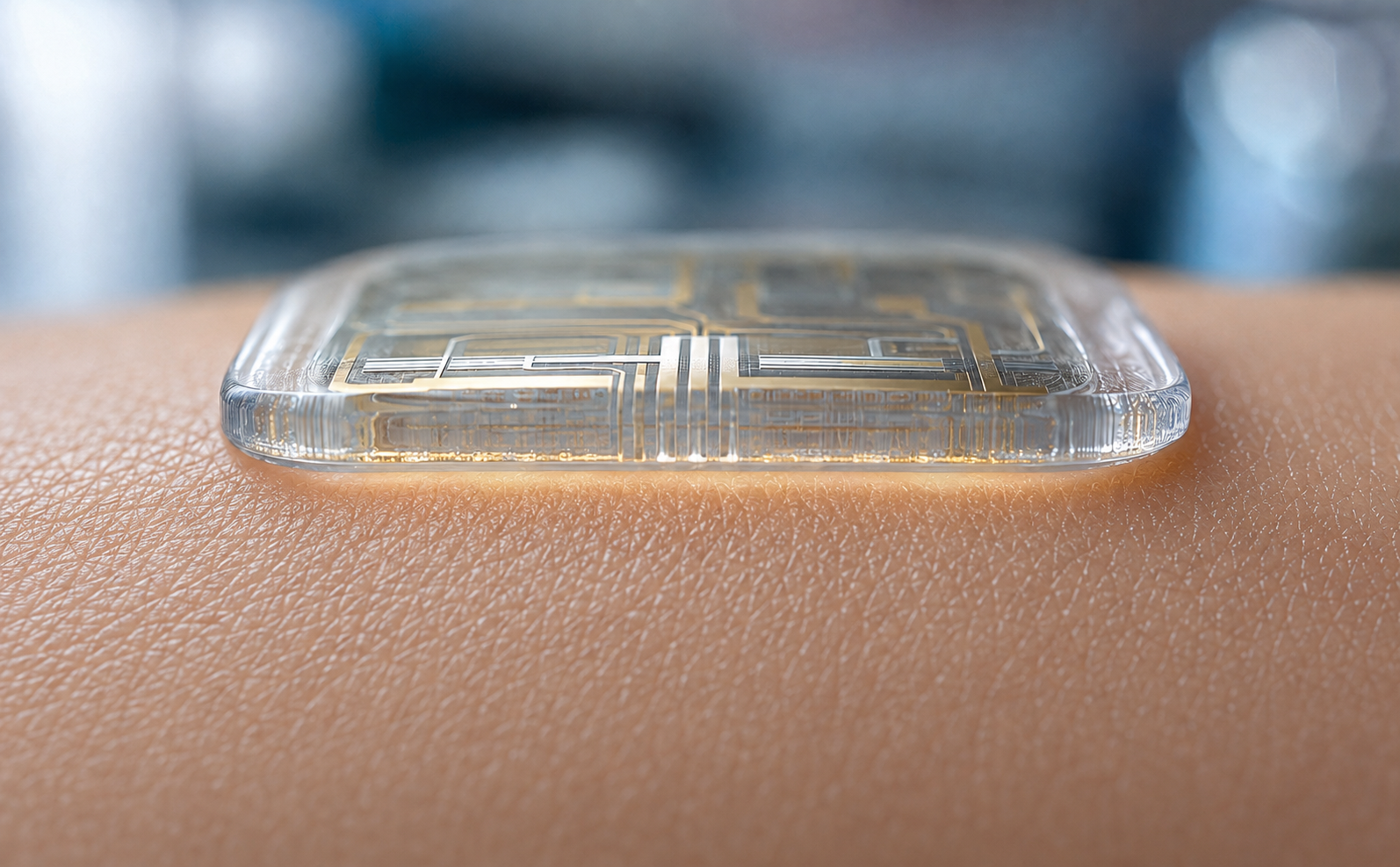
Study: In situ electrochemiluminescence microneedle device for real-time biomarker monitoring in vivo
When someone is rushed into an emergency room with chest pain, how quickly doctors identify and treat a heart attack can influence the patient’s outcome. Conventional blood tests provide measurements at individual time points rather than continuous biomarker trends, making rapid changes harder to follow.
A new study published in the journal Nature Communications describes a wearable device that uses microneedles to measure heart-related proteins in the interstitial fluid beneath the skin at repeated intervals, potentially enabling future systems to help clinicians track cardiac biomarker changes and detect patterns associated with serious cardiac events if the approach is validated in humans.
Wearable health monitoring
Wearable health technology is increasingly viewed as a potential complement to conventional blood testing, as it can track the body's condition over time rather than relying on occasional snapshots that involve invasive testing.
Interstitial fluid, which surrounds the cells under the skin, contains many of the same biological markers found in blood and is considered a promising, less invasive source for monitoring disease. Microneedle patches allow this fluid to be sampled through the outer layers of the skin with minimal invasiveness. However, existing biosensors can face modality-specific limitations. Electrochemical methods may experience interference and reproducibility problems, fluorescence systems often require bulky optics, and some interstitial fluid collection approaches depend on an external force to draw fluid from the skin.
Electrochemiluminescence pairs light emission with electrical control and offers high sensitivity, but commonly used ECL coreactants can be toxic, while conventional emitters may provide insufficient brightness and biocompatibility for use in living tissue. These limitations highlight the need for a safe, bright, biocompatible platform to track protein biomarkers in real time.
The current study
In the present study, the researchers designed a bracelet-style wearable system that paired a disposable microneedle patch with a reusable circuit board, a miniature electrochemical unit, and a light-sensing photomultiplier tube.
The microneedles, each about 1 mm long, were produced through three-dimensional printing and coated with thin coatings of gold to form working and counter electrodes, and silver to form the reference electrode. The team used a material called HOF-101, a hydrogen-bonded organic framework chosen for its brightness and preliminary evidence of low cytotoxicity, as the emitter, along with gold nanoparticles and chitosan to support the sensing layer.
The study used Y-shaped probes formed from two aptamer-containing strands, an auxiliary probe, and a ferrocene-tagged quenching strand to detect specific proteins. In their resting state, these probes suppress the light signal from HOF-101. When a target protein, such as cardiac troponin I (cTnI), binds to the aptamer arms, the quenching strand is released, allowing the light signal to recover in a concentration-dependent manner.
The sensor was first tested in artificial interstitial fluid to assess its sensitivity, specificity, repeatability, and stability. It was then evaluated in vivo using rat and pig models. Fifteen male rats, with five in each group, underwent surgery to block a coronary artery, a sham surgery, or no procedure, and wore the patches on the abdomen while signals were recorded at 30-minute intervals over 6 hours.
A similar design was tested in one 30 kg Bama pig, whose skin and cardiac anatomy more closely resemble those of humans, with the same animal monitored for 16 hours while healthy and again for 16 hours after a heart attack was induced the following day. Blood samples were collected at matching time points throughout the observation period and analyzed by ELISA, allowing temporal biomarker trends to be compared with those in serum.
Key insights
The study found that the wearable microneedle device could detect cTnI at very low concentrations in artificial interstitial fluid, while serial measurements distinguished rats with experimentally induced heart attacks from control animals. In laboratory testing, the sensor detected cTnI across a broad range, from 100 femtograms to 10 nanograms per milliliter, with a detection limit of 21.3 femtograms per milliliter. HOF-101 also generated an electrochemiluminescence signal approximately 87 times as intense as a conventional ruthenium-based ECL emitter.
Furthermore, in vitro testing showed stable repeatability for the first 12 days and relative signal stability across 25 consecutive scanning cycles. The signal was not disturbed by other acute myocardial infarction-related biomarkers at concentrations up to 100 times that of cTnI, supporting analytical specificity in artificial interstitial fluid.
In rats that had undergone surgery to block a coronary artery, the cTnI signal showed a significant upward trend after approximately 3 hours, with the temporal pattern broadly consistent with serum ELISA results from a subset of three rats with induced heart attacks. Rats given a sham operation or no surgery showed comparatively lower readings over the same period, and the biomarker profiles of the groups were statistically distinguishable as early as 1 hour after the procedure. Separate cardiac troponin T measurements also differentiated the heart attack group from the controls.
When tested on the single pig, whose skin and cardiovascular anatomy more closely resemble those of humans than those of rodents, the device recorded a relatively stable signal while the animal was healthy and a rising signal during the first 6 hours after a heart attack was induced in the same animal. The rate of signal change then remained relatively stable through the remainder of the 16-hour observation period.
However, the study did present some limitations. The animal sample was small, with five rats per group and only one pig, which served as its own healthy comparison, and the rat experiments were not blinded. Moreover, the device currently relies on a wired connection that allows only one biomarker to be tracked per session. Additionally, the mechanical stability of the skin interface during movement may affect accuracy, and the device has not yet been tested in human subjects. The animal comparisons evaluated biomarker trends and did not establish clinical sensitivity, specificity, or diagnostic accuracy.
Conclusions
Overall, the study demonstrated that a microneedle-based wearable sensor could track a key heart attack biomarker serially in living animals, while laboratory testing showed high analytical sensitivity, and the animal biomarker trends were broadly consistent with serum ELISA results.
Although the device has been tested only on animal models so far, the approach suggests a path toward wearable devices that could support frequent cardiac biomarker monitoring, although human studies must establish its safety, diagnostic accuracy, response time, and clinical value.
Source:
- Xiong, H., Zhu, C., Ashraf, G., Guo, X., Liu, L., Wang, H., Chen, H., Weng, W., Shen, H., Kong, J., & Fang, X. (2026). In situ electrochemiluminescence microneedle device for real-time biomarker monitoring in vivo. Nature Communications, 17(1), 6334. DOI: 10.1038/s41467-026-70686-8, https://www.nature.com/articles/s41467-026-70686-8
 Crucial X Chromosome Protein Protects Female Hearts From Fatal Failure