Scientists have engineered fluorescent molecules that enable the visualization of DNA organization within living cells with unparalleled resolution and, in fixed cells, nearly the width of the double helix itself.
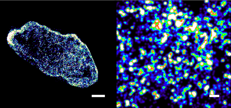 DNA inside a healthy cell (left) and a cancer cell (right) from human bowel tissue, imaged at a scale of billionths of a meter. In the healthy cell, DNA is gathered into tight, distinct packages. In the cancer cell, it has come loose and spread out. Image Credit: Aiping Wang/Centro de Regulación Genómica/Guangdong Provincial People’s Hospital
The group applied these fluorescent probes to sections of bowel tissue obtained from three cancer patients. These are standard wax-embedded specimens, which are the common method hospitals use to store patient samples.
In the cancerous growths, the DNA was distinctly less compact and more dispersed than in the adjacent healthy tissue.
Download a free copy of this page here!
Previous research indicates that DNA gradually unpacks as cancer progresses, and the scientists propose that the degree of a cell's DNA folding could ultimately serve as an indicator of tumor advancement or its virulence.
Physicians currently analyze these biopsy types visually, utilizing a staining technique over a century old. This breakthrough, detailed in the journal Molecular Cell, raises the prospect that they might someday also examine how DNA occupies three-dimensional space inside cells as an additional clue for cancer diagnosis and treatment.
With the same dye we can do two very different things. In a living cell we can watch DNA moving, which tells us how chromatin, the natural state of DNA in cells, behaves. In a preserved cell, we can zoom in until we are almost at the scale of the DNA molecule itself. Combining both approaches helps us see one of the main layers of control in human biology in unprecedented resolution.
Pia Cosma, Study Senior Author and Research Professor, ICREA
Within every human body cell, two meters of DNA are compressed into an extremely confined space. The extent of its intricate folding dictates which specific genes are activated and which ones stay inactive.
Nearly all visualizations of DNA coiling originate from non-viable cells. The powerful microscopy methods used to monitor individual DNA elements within cells typically require exposing samples to harsh chemicals and intense laser illumination, which living cells cannot withstand.
Researchers from the Centre for Genomic Regulation (CRG) in Barcelona, the City University of Hong Kong, and the Guangdong Provincial People's Hospital (Guangdong Academy of Medical Sciences), Southern Medical University, discovered a novel approach to address this issue through the development of innovative fluorescent probes.
Designated HoTs, these dyes independently traverse within viable cells and bind to DNA. The probes are engineered to blink intermittently, displaying an on-off flickering pattern akin to fairy lights. The scientists evaluated them using live human skin cells and cultured HeLa cancer cells in a lab environment.
If all dyes illuminate simultaneously, microscopes would capture a blurred image. By flashing sequentially, an advanced microscope precisely determines the position of each probe. Following the acquisition of thousands of rapid images, a computer program constructs a visualization that is 10 times clearer than that of standard microscopes.
The key challenge was to design fluorophores with the right blinking behavior for super-resolution imaging.
Hongyan Sun, Study Co-Senior Author and Professor, City University of Hong Kong
A conventional microscope blurs anything closer than approximately 200 nanometers. These novel probes enabled the scientists to employ a sophisticated microscopy technique known as STORM to visualize DNA at a 20-nanometer resolution inside live cells.
The researchers achieved even greater magnification with cells that were chemically fixed and preserved, locking their structures in place. By applying a method called MINFLUX to fixed laboratory cells, they precisely localized individual dye molecules to within three billionths of a meter, or three nanometers, nearly the width of a DNA strand.
The diameter of the DNA double helix is approximately two nanometers, so three-nanometer localization precision brings us remarkably close to the physical scale of the DNA molecule itself.
Aiping Wang, Study First Author, Southern Medical University
The probes also functioned in non-human tissues. The research group applied them to sections of zebrafish eye, an organism capable of regenerating damaged retina. It is hypothesized that retinal regeneration in these creatures involves cells relaxing their DNA to regain plasticity.
In a distinct experiment, the investigators demonstrated an AI's capability to interpret these images. They utilized AINU (“AI of the Nucleus”), a program introduced by the same group in 2024 that analyzes super-resolution cellular DNA images to identify patterns imperceptible to the human eye.
Trained on novel live-cell images, AINU accurately differentiated skin cells from stem cells 96–98% of the time.
The two cell types possess identical DNA, yet exhibit distinct folding patterns, and the AI was able to discern this distinction. When AINU was initially developed, it could only examine cells that had been fixed and preserved; its integration with the novel dyes allows it to operate on live cells.
This is the same principle the team anticipates could someday be applied to distinguish cancerous tissue from healthy tissue, or to identify the most viable stem cells to accelerate progress in regenerative medicine.
However, fluorescent molecules have clear limitations, as they envelop the entire DNA structure rather than targeting a specific gene. The sharpest images necessitated fixed cells, not live ones, though the group states it is addressing this in ongoing follow-up research, and MINFLUX offers the capability to image these probes within live cells.
Source:
Journal reference:
Wang, A., et al. (2026) HoT auto-blinking probes enable real-time, super-resolution chromatin imaging in live cells and tissues. Molecular Cell. DOI:10.1016/j.molcel.2026.08.010. https://www.sciencedirect.com/science/article/abs/pii/S1097276526005575?via%3Dihub.
 Three-Dimensional Genome Architecture Actively Dictates Phenotypic Novelties in Octopuses