Scientists at the MRC Laboratory of Medical Sciences (LMS) have devised a novel method for improving the accuracy of luminescent biological reporters, which enables researchers to monitor gene expression in living plants for days, weeks, and potentially months or even years. This technique could help scientists better understand how plants respond to diseases, pests, and environmental stress, while also providing opportunities for related applications in animal and human disease models.
 Image credit: AnaLysiSStudiO/Shutterstock.com
Published in Nature Plants, the research tackles a persistent issue in autoluminescence imaging, a method allowing organisms to produce their own light independent of additional chemicals or specialized lab apparatus.
Separating Biology from Background Noise
Researchers frequently employ light-producing reporter systems to visualize the activation and deactivation of genes. In autonomously functioning luminescent systems, the emission of light is influenced by various elements, including not only gene expression but also fluctuations in metabolic processes, temperature, oxygen concentrations, and additional factors.
This complexity poses challenges in accurately establishing whether variations in luminosity genuinely represent biological processes or are merely indicative of alterations in the organism’s physiological condition.
To address this issue, scientists developed a fungal luciferase variant that emits red-shifted light and paired it with a green luminescent reporter. By assessing the proportion of the two colors instead of the total brightness, they produced a system that maintains consistency despite changing metabolism.
The method successfully takes autoluminescence from a qualitative sign into a quantitative assessment measurement, offering a more precise understanding of the processes occurring within living organisms.
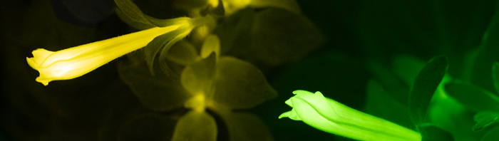
Image Credit: Light Bio
Watching Plant Defences in Real Time
Using plants, the team can observed essential defensive and immune signaling pathways. Using the developed dual-color reporters, they could watch hormone-induced reactions over prolonged durations and identifying localized immune responses near infection points, despite variations in overall brightness due to metabolic differences.
The system functioned within both fungal and bacterial bioluminescence pathways, indicating its potential for adaptation to other biological applications.
One of the biggest limitations of self-sustained luminescence has been that the brightness of the signal depends on both gene expression and metabolism. By introducing a second colour as an internal reference, we can separate those effects and obtain a much more reliable readout of what’s happening inside a living organism.
Karen Sarkisyan, Study Co-Corresponding Author Group Head, MRC Laboratory of Medical Sciences
Sarkisyan added, “This turns autoluminescence from a qualitative tool into a quantitative one. In practice, it means researchers can monitor molecular processes over long periods simply by taking photographs in the dark, without expensive equipment or invasive treatments.”
From Glowing Plants to Future Biomedical Applications
The ability to observe biological processes in a continuous and non-invasive manner could aid in the creation of “sentinel” plants that provide quantitative feedback on nutrient levels, environmental conditions, or disease through alterations in the colors of their light emissions. This technology may also assist the examining of interactions between plants and pathogens, and assessing agricultural health in more authentic field scenarios.
Although the research concentrated on plants, the researchers consider the potential wider applications. Looking into the future, the group is investigating methods to modify comparable techniques for application in animal tissues and disease simulations.
This advancement may ultimately allow researchers to observe hormone functions, gene regulation, and disease-associated mechanisms over prolonged durations using straightforward imaging techniques.
Source:
Journal reference:
Morozov, V. V., et al. (2026) Metabolically robust ratiometric autoluminescent reporters. Nature Plants. DOI:10.1038/s41477-026-02437-y. https://www.nature.com/articles/s41477-026-02437-y.
 Scientists Pinpoint the Brain Circuit That Regulates Metabolism