Scientists have built a lab-grown human liver model that stays functional for 30 days while recreating inflammation, fibrosis, and drug-induced damage, offering a promising new tool to study liver disease and test therapies without relying as heavily on animal experiments.
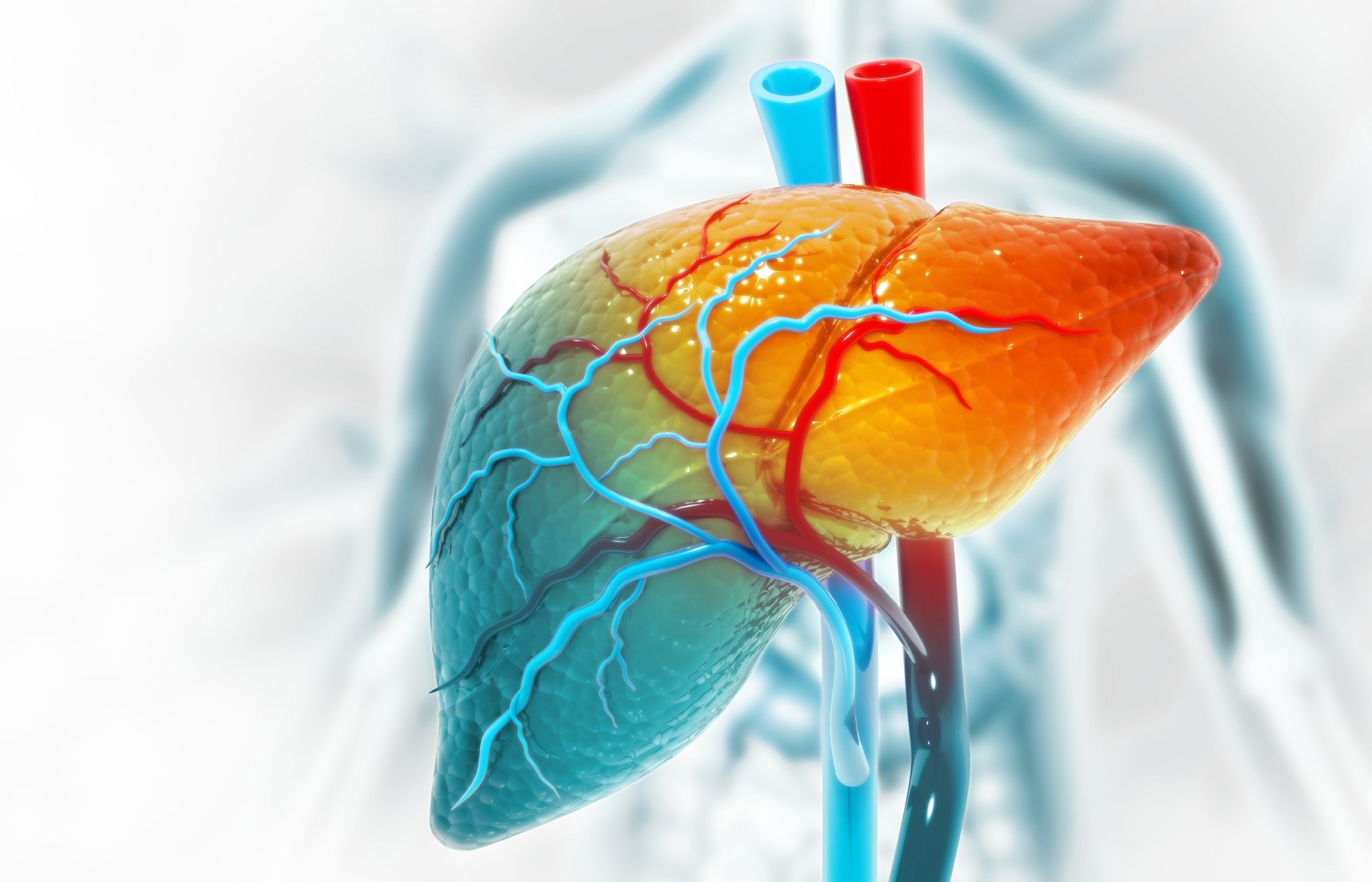 Study: A 3D bioengineered human liver for the study of acute and chronic drug-induced hepatotoxicity and fibrosis. Image credit: crystal light/Shutterstock.com
In a recent study published in Frontiers in Bioengineering and Biotechnology, researchers present a three-dimensional (3D) model that more closely replicates the human liver microenvironment than previous systems. The model remained functional for an entire month, simulating sudden liver injury and long-term diseases involving inflammation and scarring (fibrosis).
The framework also responded to dexamethasone treatment in a way consistent with its known anti-inflammatory and anti-fibrotic effects. These findings suggest that the model could help scientists test new drugs before human trials and advance personalized medicine, while reducing reliance on animal experiments.
Building A Longer-Lasting Human Liver Model
The liver performs several critical functions, including detoxifying blood, aiding digestion, and managing energy metabolism. This organ, however, is exposed to constant stress and harmful substances, which gradually contribute to liver injury. Conditions such as heavy alcohol use, unhealthy diets, infections, or even some medications can lead to liver inflammation and scarring, which may ultimately lead to liver cirrhosis or cancer.
Scientists are trying to improve the understanding of liver disease. Current models are unable to replicate long-term changes in liver function and immune interactions.
Hydrogel Scaffold Supports Month-Long Liver Function
In the present study, researchers created a durable laboratory-grown model of the human liver. To do so, they embedded human liver cells (HepaRG) along with stellate cells (LX-2) within a hydrogel. This hydrogel comprised Carboxymethyl Cellulose Methacrylate (CMCMA) and Gelatin Methacryloyl (GelMA).
The gel-like material acted as a supportive scaffold, allowing the tissue to stay functional for a month. During this period, the team tracked liver function, cell survival, fibrosis markers, and metabolic activity. Investigations included ribonucleic acid sequencing (RNA-seq), quantitative polymerase chain reaction (PCR), enzymatic assays, and high-resolution confocal imaging.
The team then simulated acute and chronic liver damage. To mimic chronic inflammatory and fibrotic changes, they exposed the lab-grown liver tissue to sustained low-dose exposure of 0.75 mM paracetamol and 10 ng/mL lipopolysaccharide (LPS). To mimic acute damage, the researchers used higher concentrations, i.e., 100 ng/mL LPS and 2.5 mM paracetamol for approximately three days.
Principal component analysis (PCA) and gene set enrichment analysis (GSEA) helped track changes in gene activity and liver function, enabling observation of disease development under controlled conditions.
The researchers then tested whether the model could respond to treatment in a biologically meaningful way. They used dexamethasone, a drug that reduces inflammation and scarring, to investigate whether it could reverse some of the liver damage in the 3D model. The team then added THP-1 human monocytes to simulate inflammatory changes in the body during liver disease. They measured the levels of inflammatory molecules, such as interleukin-1β (IL-1β), IL-6, and tumor necrosis factor-α (TNF-α), to assess these changes.
Bioengineered Liver Tissue Maintains Function For 30 Days
The engineered liver model remained functional for 30 days, a major improvement over conventional 2D systems that typically lose liver function much more quickly. During this period, the simulated liver performed crucial functions, including albumin production, drug metabolism, and the removal of toxic ammonia from the body.
The liver cells organized into stable 3D structures, which helped maintain liver activity over time. The support cells linked to liver scarring, known as hepatic stellate cells, also remained inactive unless injury was introduced. This more closely reflects aspects of human liver behavior in health and disease. Gene analyses further indicated that liver cells matured steadily between Days 10 and 30, while signals linked to cell death decreased.
Exposure to sustained low-dose LPS and paracetamol led to gradual changes in the lab-grown liver. These changes replicated those observed in chronic liver diseases in people. These included inflammation, fibrosis, fat accumulation, reduced ability to process toxins and drugs, and widespread disruptions in cellular activity and metabolism.
Over 30 days, the model showed increasing fibrosis markers. These markers included collagen type 1 alpha 1 chain (COL1A1), procollagen type III N-terminal propeptide (PIIINP), and procollagen type I N-terminal propeptide (PINP). The team also observed declining levels of important liver-function genes, including albumin, carbamoyl phosphate synthetase 1 (CPS1), and ornithine transcarbamylase (OTC).
Higher concentrations of LPS and paracetamol led to sharper declines in liver-function markers, elevated ammonia levels, and enlarged cell nuclei. These changes were accompanied by strong activation of fibrosis-related proteins, such as α-smooth muscle actin (α-SMA) and COL1A1. Fat droplets also accumulated rapidly, indicating severe metabolic stress.
The model additionally responded to treatment in a biologically relevant way. Dexamethasone reduced markers of inflammation and fibrosis while partially restoring liver-function genes to closer-to-normal levels. After monocyte addition, IL-6, IL-1β, and TNF-α increased significantly during acute and chronic injury. The findings demonstrated how immune-driven inflammation contributes to liver damage.
3D Liver Platform Advances Drug Safety Research
Researchers developed a 3D human liver model that remained functional for a month, replicated immune-related changes in disease, and simulated a treatment response. These features could help improve understanding of liver diseases and advance drug development to develop safer, more effective treatments that lower the liver disease burden worldwide.
However, the model still uses standardized cell lines and does not yet include the full range of liver cell types found in the human liver, such as endothelial and Kupffer cells. Future studies could include additional liver cell types and incorporate patient-derived cells to further improve model simulations.
Download your PDF copy by clicking here.
Journal Reference
Ferret-Miñana, A., Alcaraz, E., Horrillo, R., Ramón-Azcón, J. and De Chiara, F. (2026). A 3D bioengineered human liver for the study of acute and chronic drug-induced hepatotoxicity and fibrosis. Frontiers in Bioengineering and Biotechnology. 14:1798323. DOI: 10.3389/fbioe.2026.1798323. https://www.frontiersin.org/journals/bioengineering-and-biotechnology/articles/10.3389/fbioe.2026.1798323/full
 High Protein Levels Bypass Functional BRCA Defenses to Generate Toxic Double-Strand Genomic Breaks