By turning skin-resident memory T cells into local immune beacons, the experimental patch could offer researchers a less invasive window into difficult-to-detect immune responses.
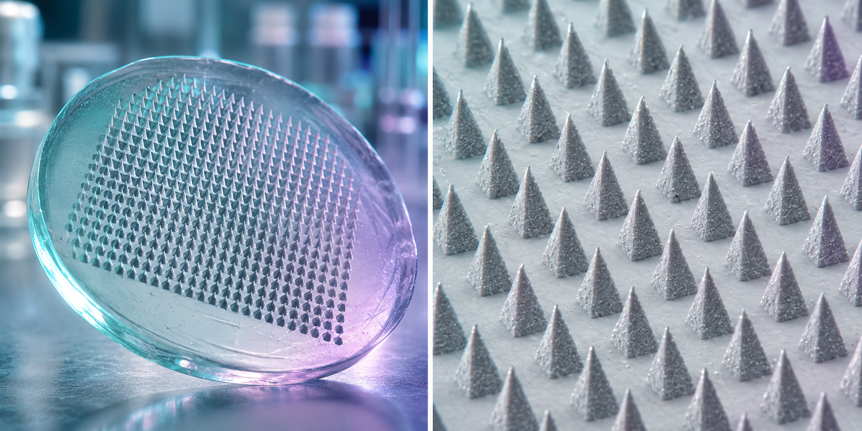
Paper: Leveraging tissue-resident memory T cells for non-invasive immune monitoring via microneedle skin patches
In a recent study published in Nature Biomedical Engineering, researchers describe the development and performance of a novel minimally invasive immune-monitoring platform that couples tissue-resident memory T cell (TRM) restimulation with hydrogel-coated microneedle (MN) skin patches.
The platform was designed to exploit the sentinel "alarm" function of skin-resident TRM cells to locally recruit rare circulating antigen-specific lymphocytes from peripheral blood into the upper dermis. An optimized alginate hydrogel coats the microneedles and captures migrating lymphocytes and interstitial fluid for immunophenotyping and proteomic analysis.
The approach was subsequently tested in murine vaccination models and in human studies assessing patch tolerability and proof-of-concept immune sampling. In mice, the platform recovered over 2-fold more live antigen-specific T cells than a 100 µL blood sample or a 6-mm punch biopsy, while being substantially less invasive than a punch biopsy, highlighting its potential for future monitoring of systemic immunity. However, the punch biopsy recovered more antigen-specific cells per unit of sampled skin area.
Background
Monitoring antigen-specific lymphocyte responses is important for evaluating immune responses to vaccination. Such monitoring may also provide insights into immunity during infection, cancer, and autoimmune disease.
Unfortunately, traditional clinical monitoring relies heavily on peripheral blood sampling, which struggles to detect antigen-specific CD8+ T cells because they can be extremely rare in circulation, accounting for approximately 0.05% of blood lymphocytes in some vaccinated populations.
While alternative tissue-sampling techniques have been developed to overcome these persistent challenges, these techniques, for example, standard 6-mm skin punch biopsies or negative-pressure suction blistering, can be invasive or painful and may cause hyperpigmentation. Suction blister samples can also contain high proportions of non-immune keratinocytes.
In parallel, while microneedle patches have been used to sample dermal interstitial fluid (ISF), patch applications without local immune stimulation typically yield negligible cell counts. However, using the rapid chemokine-driven recruitment capability of skin TRM cells to locally concentrate circulating memory T cells for minimally invasive patch recovery had not previously been established.
About the study
The researchers aimed to address this immune-sampling challenge by establishing a two-step TRM-recruitment and microneedle-capture system. The system was fabricated using biodegradable poly(L-lactide) microneedle arrays (550 µm height, 250 µm base width) coated with a swellable hydrogel matrix composed of ultra-pure, low-molecular-weight alginate (SLG20) and sucrose, crosslinked with calcium chloride (CaCl2).
The system’s performance was initially tested in preclinical murine models, specifically C57BL/6 and KikGR photoconvertible transgenic mice, which were systemically immunized with ovalbumin (OVA) plus CpG adjuvant or an SIV mRNA-lipid nanoparticle (LNP) vaccine encoding five putative SIV T-cell epitopes, including CL9. After systemic vaccination, TRM populations were established via intradermal (i.d.) antigen-plus-adjuvant administration and recalled via i.d. antigen challenge, followed by an 18-hour MN patch application approximately one week post-recall.
Subsequently, short-term patch tolerability was tested in 45 healthy volunteers. Finally, the study conducted a human proof-of-concept experiment in one participant in whom allergic contact dermatitis was induced through sensitization and re-exposure to squaric acid dibutyl ester (SADBE).
The study’s analyses included time-lapse confocal cell tracking, flow cytometry, ELISPOT assays, and Olink proteomic profiling.
Study findings
The study’s hydrogel optimization trials revealed that the high G/M ratio, low-molecular-weight SLG20 alginate enabled ~2.5-fold higher T cell migration speeds in vitro than the other alginate formulations tested. Furthermore, SLG20 and SLM20 recovered approximately twice as much ISF in vivo as the higher-molecular-weight formulations, although statistical significance varied across the pairwise comparisons.
In vivo evaluations of OVA-immunized mice showed that intradermal TRM recall increased live cell recovery 20-fold over non-TRM controls. Compared with skin containing established but unrecalled TRM cells, recall increased the recovery of CD4+ T cells by 6.5-fold, CD8+ T cells by 8-fold, and OVA-tetramer+ CD8+ T cells by 18-fold (p = 0.0002, p < 0.0001, and p < 0.0001, respectively).
Photoconversion studies in KikGR mice and systemic LFA-1 antibody blockade indicated that at least half of captured antigen-specific T cells were actively recruited from peripheral blood circulation. When evaluated against the SIV mRNA-LNP vaccine, TRM recall was found to induce a 7.5-fold increase in CL9-tetramer+ CD8+ T cell recovery (p = 0.0036).
Together, in mice, a 2 cm² MN patch yielded >2-fold more live antigen-specific T cells than a standard 100 µL blood draw or a 6-mm skin punch biopsy (MN versus blood, p = 0.0077; MN versus biopsy, p = 0.0169). Although the patch recovered more antigen-specific CD8+ T cells overall, the biopsy recovered more antigen-specific cells per unit of sampled skin area.
Finally, the human tolerability assessment showed that the platform’s 4 cm² MN patches were well tolerated across all 45 participants, causing only mild redness that resolved within one hour, with no reported bleeding, swelling, or other adverse reactions.
In the single-participant SADBE allergen recall model, MN patches primarily recovered viable CD45+ immune cells (CD4+, CD8+, NK cells, dendritic cells, and monocytes), whereas suction blisters recovered more total live cells but predominantly collected probable non-immune keratinocytes. Concurrently, descriptive Olink proteomic profiles showed increases in T-cell homing chemokines, including CXCL9, CXCL10, and CXCL11, as well as IFN-γ and other cytokines associated with T-cell activation.
However, the researchers could not directly confirm that the human cells were SADBE-specific because validated tetramers and canonical epitopes for this allergen are unavailable.
Conclusions
The present study demonstrates that coupling skin TRM cell restimulation with hydrogel-coated microneedle patches provides a promising, minimally invasive approach that could support longitudinal sampling of both tissue-resident and circulating antigen-specific immune cells. Evidence that the patches recovered circulating antigen-specific cells came from the mouse models, whereas the functional human sampling experiment involved one participant.
Larger, controlled human studies are required to establish performance across different diseases, anatomical sites, and patient populations. The hydrogel patch formulation does not require antigen or adjuvant loading and could therefore be used across different immune-monitoring applications. However, the tested procedure still depends on the presence of responsive antigen-specific TRM cells and local recall stimulation, which could, in turn, alter local immune activity.
 New CRISPR Atlas Details How Every Gene Influences Stem Cells