Researchers at Cambridge have created tiny circuits in the lab that replicate the connections between the brain and spinal cord that control human movement. Using this model, they demonstrated that damage to these connections, once thought "irreversible," might actually be reversible.
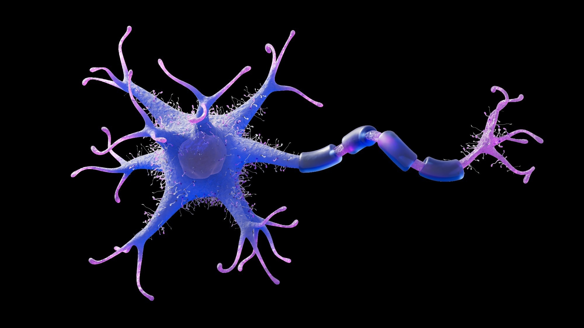 Image credit: Ursa Design/Shutterstock.com
The nerve cells, or neurons, join as humans grow from embryo to fetus to infant, facilitating information transfer between the brain and the spinal cord. The axon, the nerve fiber "cable" that sends signals to neighboring neurons to initiate muscular contractions, is an essential part of every neuron.
People eventually lose the capacity to develop axons in the central nervous system, or at the very least, this capacity is significantly reduced or slowed. This results in irreversible brain and spinal cord damage, which causes severe impairments such as the incapacity to grasp or walk. This can be a characteristic of many neurological conditions, such as multiple sclerosis or motor neuron disease, and is frequently the case for traumatic spinal cord injuries.
Researchers at the University of Cambridge created "mini brains" in 2021 by guiding human patient-derived stem cells, which are unique cells capable of differentiating into most human cell types, to develop into pea-sized brain "organoids." These organoids were three-dimensional representations of portions of the human cerebral cortex. These were used by the team to illustrate molecular mechanisms underlying motor neuron disease and potential preventive measures.
In a recent study published in Cell Reports, Dr. Lakatos's team went one step further, using organoids to recreate these tissues and create a miniature version of the interconnected human brain and spinal cord system.
The brain and spinal cord organoids were kept separate by the researchers, since the brain and spinal cord tissues in the human body are distinct yet connected by axons. They saw that nerve fibers from the brain tissue extended over the opening to join the spinal cord, creating a functional circuit that could even trigger the contraction of small muscle groups.
After growing this human system in a dish for almost a year, they discovered that the axons could recover from injury until about day 150, or the mid-trimester of pregnancy, at which point their growth was significantly hindered.
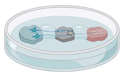 Illustration of the model, with the brain organoid, spinal organoid, and muscle tissue (left to right). Image Credit: András Lakatos lab
George Gibbons from the Department of Clinical Neurosciences said: “Neurons taken from less mature organoids regrew long fibres after injury, but those from more mature organoids showed a sharp drop in their ability to regrow. In other words, poor regeneration is built into human neurons as they mature in the central nervous system.”
They discovered a network of genes that functions as a "switch" limiting the ability of axons to develop while the neurons mature to create connections (synapses) by analyzing the gene expression, a measure of how active the genes are, in neurons that connect the brain and the spinal cord. Amazingly, axon growth was restored when important network regulators were blocked.
Lynestrenol, a hormone medication approved for the treatment of specific menstrual diseases and as a contraceptive, was found as a candidate by the researchers after they searched a database of chemical compounds for those that act on the genes in this network. They discovered that this medication greatly increased axon regeneration when they tested it on injured neurons.
Project leader Dr. András Lakatos said: “When the brain and spinal cord are damaged, the nerve fibres that carry movement signals from the brain to the spinal cord rarely grow back. That’s why paralysis is usually permanent. But we didn’t know exactly when the ability of axons to regenerate becomes limited. Our model provides a good indication that this block happens during development, and it can still be reversed after this point."
Lynestrenol itself may not be the answer to spinal cord repair, but it shows us that, in principle, it should be possible to directly target human neurons and regenerate their axons. Although we still need to show that this strategy will also help to re-establish appropriate connections between the brain and spinal cord cells, this gives us hope that one day we may be able to treat conditions previously thought untreatable.
Dr. András Lakatos, Department of Clinical Neurosciences, University of Cambridge
One of the most important tools for understanding human biology is the use of organoid models. While animal models such as mice and rats have long been valuable for biological research due to their similarities to humans, key differences between species can limit the insights they provide. Organoids, generated from human stem cells, offer a more physiologically relevant model that more closely replicates human biology.
Much of what we know about nerve regeneration comes from rodents, whose neurons behave differently from human neurons. Our sophisticated organoid models help bridge the knowledge gap from animal models to what we see in patients. They are also an important contribution to efforts to reduce the use of animals in research.
Dr. András Lakatos, Department of Clinical Neurosciences, University of Cambridge
The use of organoids, sometimes known as "mini organs," to simulate human biology and illness is growing. Researchers at the University of Cambridge use them for a variety of purposes, including modeling the early stages of pregnancy, understanding Crohn's disease in youngsters, and repairing damaged livers.
Today, we are entering a new era of hope and possibility for the 15 million people worldwide living with a spinal cord injury. The next five years present an unprecedented opportunity to change what’s possible for people living with spinal cord injuries. Breakthrough therapies are nearing clinical reality and frontier technologies are creating bold new pathways toward repair and recovery.
Louisa McGinn, Spinal Research Chief Executive, University of Cambridge
Source:
Journal reference:
Gibbons, G. M., et al. (2026) A human corticospinal organoid-slice connectoid model informs enhancer strategies for post-injury axon regrowth. Cell Reports. DOI: 10.1016/j.celrep.2026.117399. https://www.cell.com/cell-reports/fulltext/S2211-1247(26)00477-8.
 Female Hearts Rely on Molecular Safeguard to Avoid Failure