One of the earliest casualties of aging is skeletal muscle. As muscles begin to deteriorate, they can become weaker and more scarred, with fat gradually accumulating within the tissue. At the same time, fast-twitch muscle fibers, which power rapid and forceful movements, are progressively lost.
 Image credit: longwan/Shutterstock.com
A study team from Kyushu University’s Faculty of Agriculture has recently discovered a molecule that might improve and safeguard one of the body’s primary signals for muscle healing. The results were released in Scientific Reports.
The study focuses on hepatocyte growth factor (HGF), a molecule that acts as a wake-up signal in skeletal muscle. In healthy tissue, HGF lies dormant within the supportive network surrounding muscle fibers. When muscle is damaged or mechanically stimulated, HGF is released and binds to c-met receptors on satellite cells, the resident stem cells of skeletal muscle. This activates the cells, allowing them to proliferate, differentiate, and contribute to muscle repair.
But this process can be hampered by age. Previous research by the team demonstrated that HGF goes through a chemical process known as nitration, in which a nitro group is added to two particular protein sites, Y198 and Y250, the area that HGF uses to bind c-met. Like a rusty key that no longer fits its lock, HGF can no longer dock with the receptor once it has been nitrated. This is believed to be the primary cause of poor regeneration and age-related muscle atrophy.
HGF is not necessarily missing as we age. Rather, it can be chemically altered after it is made. That led us to wonder whether a compound with strong antioxidant capacity might protect HGF, either by preventing nitration or by compensating for the functional loss it causes.
Ryuichi Tatsumi, Professor, Faculty of Agriculture, Kyushu University
The group used glutathione trisulfide (GSSSG) and lipoic acid trisulfide (LASSS), two sulfur-based compounds with potent antioxidant activity. Both are members of the group of molecules known as trisulfides, which have three sulfur atoms connected in succession. Recently, their unique sulfur chemistry and redox characteristics have drawn interest for pharmaceuticals.
Initial experiments showed that both GSSSG and LASSS suppressed HGF nitration at Y198 and Y250. However, because receptor-binding activity was not fully restored, the researchers increased the molar ratio of HGF to trisulfide from 1:4000 to 1:8000.
The results were unexpected. At the higher ratio, HGF's binding affinity for c-met more than doubled compared with untreated HGF, while also becoming more resistant to nitration-induced dysfunction, particularly at Y198. Notably, this effect was observed only with LASSS; GSSSG did not produce the same enhancement.
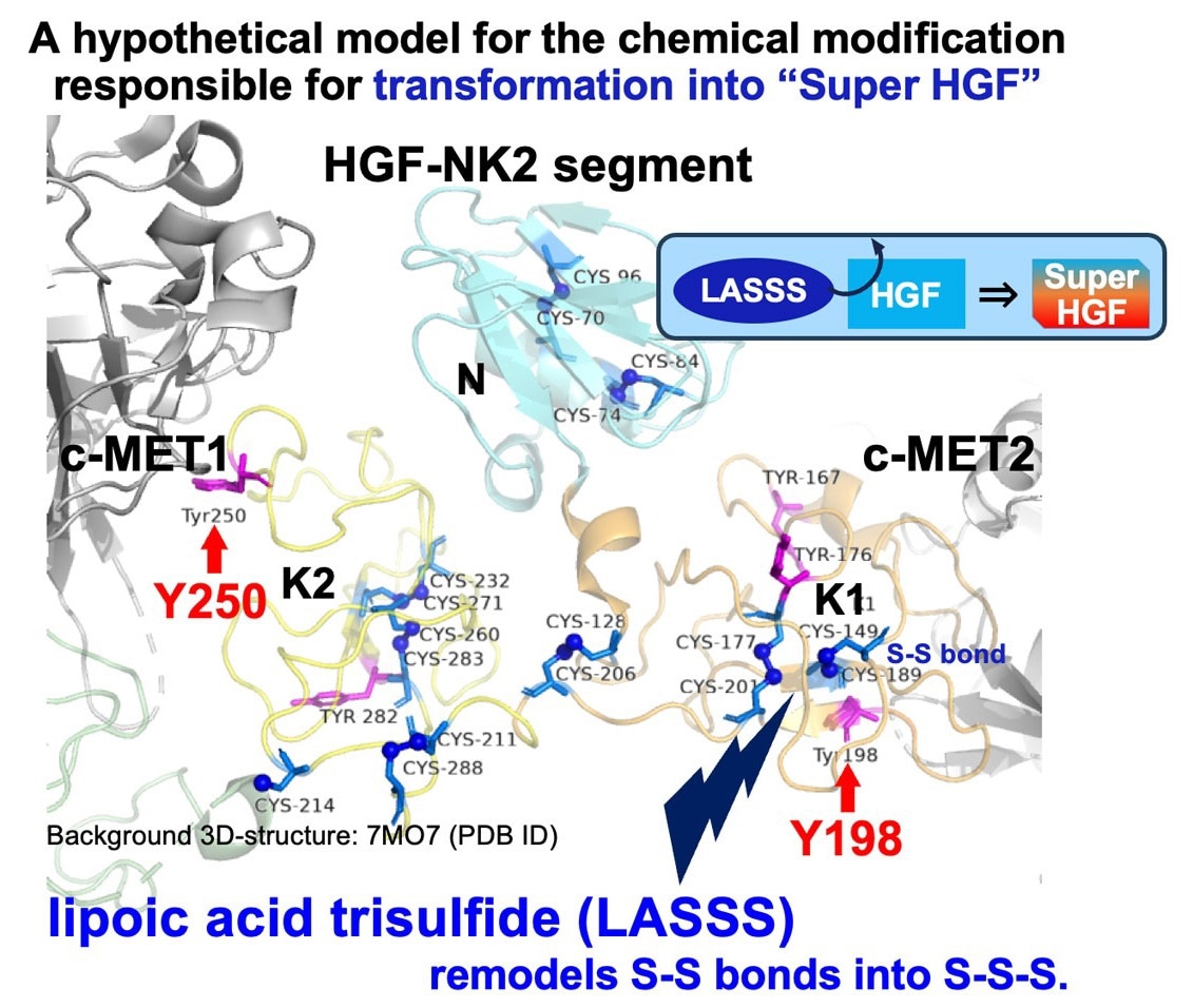 Lipoic acid trisulfide (LASSS) may turn HGF into “Super HGF”. HGF is a key activator of muscle stem cells that normally resides in the extracellular matrix, but becomes dysfunctional with age due to nitration. Researchers show that interaction with lipoic acid trisulfide (LASSS) can convert HGF into “Super HGF,” a form with enhanced receptor-binding affinity and resistance to nitration-induced dysfunction. Disulfide bonds may be potential targets for LASSS-mediated remodeling into trisulfide bonds. Image Credit: Kyushu University
This exceeded our expectations. We knew trisulfides had diverse biological functions, but we never expected that simply mixing HGF with LASSS would produce such a striking effect. What this tells us is that LASSS does more than simply neutralize reactive molecules. It may interact directly with HGF and induce a subtle structural change, creating an enhanced ‘Super HGF’ form that binds c-met more strongly and resists nitration.
Ryuichi Tatsumi, Professor, Faculty of Agriculture, Kyushu University
To investigate whether the protective effects of LASSS extended beyond cell-based experiments, the team tested the compound in mice experiencing muscle atrophy induced by tail suspension. Animals that received LASSS treatment showed markedly lower levels of protein nitration than untreated mice, whereas GSSSG again failed to provide any measurable benefit. These results suggest that LASSS retains its protective properties under more physiologically relevant conditions, although additional studies in older animals will be needed to assess its long-term safety and therapeutic potential.
The researchers say their findings could eventually inform new approaches to supporting muscle regeneration in situations where muscle activity is reduced, including aging and prolonged immobilization. They also propose that the HGF-protective effects of LASSS may not be limited to humans and could extend to other mammals, including companion animals. If confirmed, such benefits could help sustain mobility, independence, and overall wellbeing later in life.
Source:
Journal reference:
Zushi, K., et al. (2026) Enhanced HGF with increased receptor affinity and nitration-dysfunction resistance through interaction with lipoic acid trisulfide. Scientific Reports. DOI: 10.1038/s41598-026-60835-w. https://www.nature.com/articles/s41598-026-60835-w.
 Single-Molecule Optical Tweezers Visualize Real-Time Dynamic Protein and DNA Interactions