Tissue engineers are developing methods to produce living organs and tissues from cells in order to replace diseased and damaged ones in the body. Artificial muscles, livers, kidneys, skin, and other tissues have been successfully grown by scientists. However, carefully structured networks of blood vessels, some of which can be finer than a human hair, have not been reliably engineered.
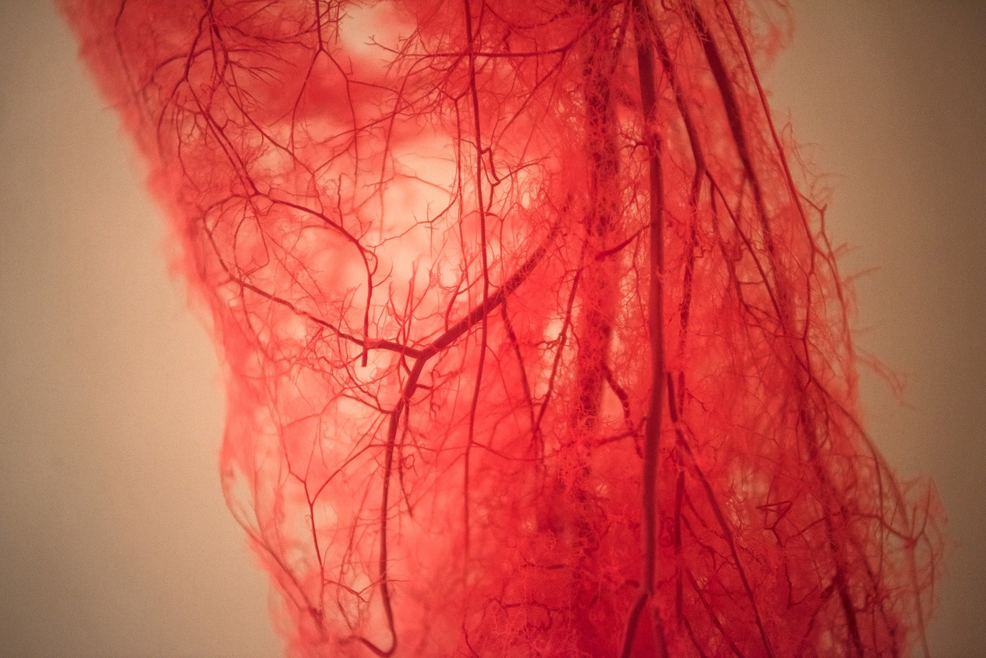 Image credit: Captain Wang/Shutterstock.com
Artificial tissues, regardless of how realistic they are, cannot operate without a vascular network to supply nutrition. But by mechanically stretching blood vessels, MIT engineers have discovered a way to design and development them.
The group has created a human "blood vessel on a chip," which consists of a core artery consisting of human endothelial cells encased in a gel with a tiny magnet. Using an external magnet to move the magnet implanted in the gel, the researchers jostled the gel back and forth to observe how the major artery reacted.
They discovered that the artery began to grow new, smaller capillaries. By altering the direction in which the artery is stretched or jostled, researchers might reroute the developing new arteries. Furthermore, depending on how much the artery was stretched, the number of additional vessels that grew also varied.
Their findings, which were published in the Proceedings of the National Academy of Sciences, provide researchers with a fresh approach to creating artificial blood arteries and controlling their growth patterns.
Healthy tissues depend on organized blood vessel networks, but state-of-the-art protocols don't enable fabricating such networks within engineered tissues. The ability to program blood vessel growth with physical cues may enable reproducible and scalable fabrication of engineered tissues that can be implanted in the body to restore function after debilitating disease or injury.
Ritu Raman, Study Co-Lead Author and Associate Professor, Mechanical Engineering, Massachusetts Institute of Technology
“Moving is Good”
Blood vessels are difficult to produce using traditional fabrication methods. While 3D printers can create vessels on the size of major arteries and veins, the technology is insufficiently accurate to print complicated networks of much finer, thread-like capillaries.
Scientists have had some success creating blood arteries from single cells by culturing them in Petri dishes with nutrients and growth factors. However, it is still difficult to manage how and where they develop.
You can try to pattern chemical cues, like growth factors, to direct where vessels grow, but you can’t do this very precisely. We thus need other types of patternable cues that can help us build tissues with organized vessels.
Ritu Raman, Study Co-Lead Author and Associate Professor, Mechanical Engineering, Massachusetts Institute of Technology
She and her students wondered whether they might use a technique they had previously created to produce artificial muscles and nerves to build and regulate new blood vessels. In earlier research, the group created a tiny chip that contained a gel that was enriched with growth hormones and nutrients.
After incorporating a tiny magnet into the gel, they covered its surface with living muscle or neuronal cells. The embedded magnet and the cell-covered gel were then pulled back and forth by manipulating an external magnet. This research demonstrated that the cells' growth was directly impacted by mechanical "exercise," which involved pushing the cells back and forth.
The researchers employed a similar arrangement in their recent study to test their ability to develop and regulate new blood vessels.
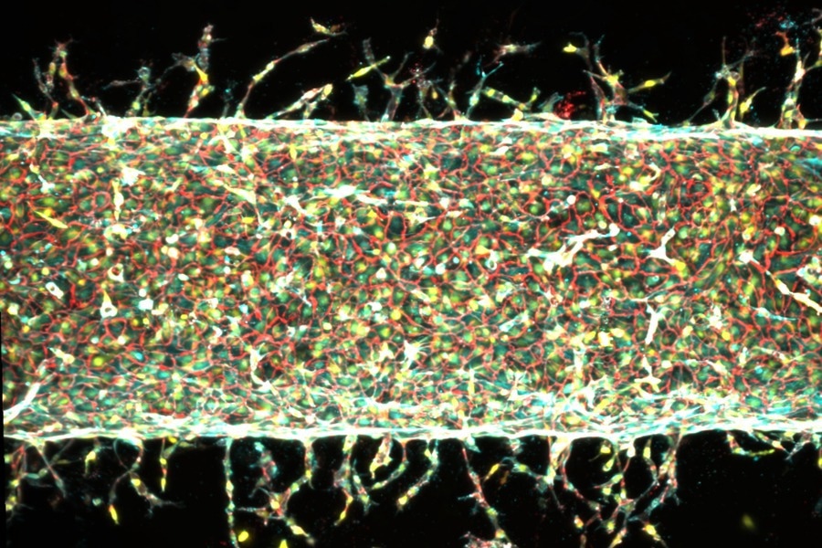 With mechanical stretching, MIT engineers can control how artificial arteries sprout new capillaries. Image Credit: Courtesy of the researchers, MIT
Smaller than a postage stamp, the researchers constructed a "blood-vessel-on-a-chip" and filled it with a nutrient-rich gel that included a magnet. They created a hollow channel by poking a narrow tube lengthwise through the gel.
They then covered the channel with living endothelial cells, which spontaneously proliferate and fuse to form blood vessels in the body. The cells began to sprout new, capillary-like capillaries in the gel after they assumed the geometry of the channel.
Placing the device under a motorized stage fitted with small, suspended magnets, the researchers moved the magnets back and forth in different directions, and by various degrees, and observed whether and how blood vessels sprouted from the central artery in response.
The main takeaway is: Stretching the blood vessel back and forth seems to enhance the number of new capillaries that grow.
Ritu Raman, Study Co-Lead Author and Associate Professor, Mechanical Engineering, Massachusetts Institute of Technology
The major artery would develop several additional arteries at random points throughout its length if it were just left alone in the gel. However, a lot more vessels appeared when the artery was disturbed.
Numerous additional arteries sprang from the main artery when the scientists used the magnets to stretch the gel back and forth by 5% of its overall diameter. Fewer vessels emerged when they extended by 15%, but the ones that did became longer.
Additionally, the new vasculature responded by taking turns and following the team's mechanical stimulation pattern when the researchers changed the direction of stretching.
“We’re finding that moving is good, which is always the takeaway of everything we do in our lab,” Raman says. “Mechanical forces play an important role in our bodies. That means that if you want to grow more or less vessels, or shorter or longer vessels, or vessels in certain directions, we now know how to do that.”
A Gatekeeping Gene
To understand why blood vessels expand in reaction to mechanical pressures, the researchers went one step further. They used gene editing and the function of a specific gene, Piezo1.
Raman had just gone to a talk given by Ardem Patapoutian, a molecular biologist. Patapoutian's discovery of ion channels in cell membranes that open and close in response to mechanical pressure earned him the 2021 Nobel Prize in Physiology or Medicine. These channels, PIEZO1 and PIEZO2, serve as a cell's gatekeepers, regulating what enters and exits the cell. Patapoutian discovered that the corresponding genes, also known as PIEZO1 and PIEZO2, control both kinds of channels.
Following his talk, Raman presented Patapoutian with the experimental findings from her lab, which demonstrated a link between mechanical stimulation and blood vessel expansion. Patapoutian suggested that the PIEZO1 channel may be the reason; by manually exercising the central artery, Raman would have been encouraging the opening of ion channels in the artery's cells, which would have led to the growth of additional blood vessels.
Raman tested this idea by suppressing the PIEZO1 gene in endothelial cells. When these edited cells were mechanically stimulated, far fewer new blood vessels formed, indicating that mechanically driven vessel growth depends on PIEZO1 activation.
The team intends to use the protocol to develop ordered networks of vessels to supply artificial organs and tissues now that they have figured out how to generate and regulate blood vessel growth.
“We are now investigating how precisely patterning blood vessel growth can help improve muscle function,” notes co-author Jessica Shah.
Traveling Through Our Engineered Blood Vessels
Video constructed from a 3D high-resolution microscopy image of engineered blood vessel tissue made by MIT engineers, showing a fly-through of a central artery and new capillaries that sprout from the artery in response to mechanical stimulation. Video Credit: Massachusetts Institute of Technology
Source:
Journal reference:
Kheiri, S., et al. (2026) 4D force patterning enables spatial control of angiogenesis. Proceedings of the National Academy of Sciences. DOI: 10.1073/pnas.2532667123. https://www.pnas.org/doi/10.1073/pnas.2532667123.
 Microbe Retains Self-Replication Machinery While Discarding Almost All Metabolic Pathways